


Call us
301-363-4651 (Available 9 a.m. to 5 p.m. CST from Monday to Friday)
Skin and connective tissue disorders are a group of diseases that affect the skin, muscles, bones, blood vessels and other connecting tissues. These diseases may be caused by genetics, immune abnormalities, environmental factors, or other unknown factors. Some common skin and connective tissue diseases include systemic lupus erythematosus (SLE), rheumatoid arthritis (RA), dermatomyositis, dry syndrome (SS), and systemic sclerosis.
In recent years, progress is being made in drug development for skin and connective tissue diseases, some of which are listed below:
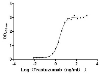
Measured by its binding ability in a functional ELISA. Immobilized HER2 at 2 μg/ml can bind Trastuzumab, the EC50 is 2.179-2.825 ng/ml.
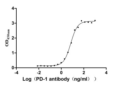
Measured by its binding ability in a functional ELISA. Immobilized PD-1 at 2 μg/ml can bind Anti-PD-1 recombinant antibody, the EC50 of human PD-1 protein is 6.087-7.854 ng/ml.

Measured by its binding ability in a functional ELISA. Immobilized human CD20 at 2 μg/ml can bind Anti-CD20 recombinant antibody (CSB-RA015007A1HU), the EC50 is 3.243-7.085 ng/mL
-AC1.jpg)
Measured by its binding ability in a functional ELISA. Immobilized Human IL17A at 2 μg/ml can bind Anti-IL17A recombinant antibody (CSB-RA624104MA1HU), the EC50 is 1.818-2.170 ng/mL.

Measured by its binding ability in a functional ELISA. Immobilized PD-L1 at 2 μg/ml can bind Anti- PD-L1 mouse monoclonal antibody (CSB-MA878942A1m, antigen from E.coli), the EC50 of human PD-L1 protein is 1.252-1.653 ng/mL

Human EGF protein captured on COOH chip can bind Human EGFR protein, his and Myc tag (CSB-MP007479HU) with an affinity constant of 11.9nM as detected by LSPR Assay.

Measured by its binding ability in a functional ELISA. Immobilized Human IL4 (CSB-MP011659HU) at 2μg/mL can bind biotinylated Human IL4R,the EC50 is 12.68-14.23 ng/mL.
Measured in cell activity assay using U937 cells, the EC50 for this effect is 190.2-298.6 ng/ml.

CT26/Human ROR1 Stable Cell Line
CSB-SC020067HU
Untransfected CT26 cells (green line) and transfected Human ROR1 CT26 stable cells (red line) were stained with anti-ROR1 antibody (CSB-RA020067A1HU) (2µg/1*106 cells), washed and then followed by FITC-conjugated anti-Human IgG Fc antibody and analyzed with flow cytometry.

Overlay Peak curve showing HepG2 cells stained with CSB-RA264109A0HU (red line) at 1:50.
Untransfected HEK293T cells (green line) and transfected Human ENPP3 HEK293T Stable cells (red line) were stained with anti-ENPP3 antibody (2µg/1*106 cells).
Overlay Peak curve showing Hela cells surface stained with CSB-RA003995MA1HU (red line) at 1:800.
Overlay Peak curve showing Hela cells surface stained with CSB-RA878942MA1HU (red line) at 1:100.
Tumour