


Call us
301-363-4651 (Available 9 a.m. to 5 p.m. CST from Monday to Friday)
Non-neoplastic blood and lymphatic disorders include a range of conditions that affect blood components and lymphatic system function, such as anemia, coagulation disorders, platelet abnormalities, and myelodysplastic syndromes. These disorders are treated in a variety of ways, including medications, blood transfusions, and bone marrow transplantation. In recent years, significant progress has also been made in the development of drugs for these non-neoplastic hematologic and lymphatic disorders.

Measured by its binding ability in a functional ELISA. Immobilized human CD20 at 2 μg/ml can bind Anti-CD20 recombinant antibody (CSB-RA015007A1HU), the EC50 is 3.243-7.085 ng/mL
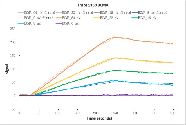
Human TNFSF13B protein Fc tag (CSB-MP897523HU1) captured on COOH chip can bind Human BCMA protein Fc tag (CSB-MP023974HU1) with an affinity constant of 39 nM as detected by LSPR Assay.
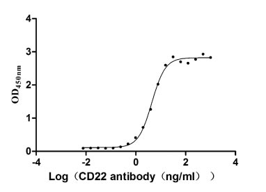
Measured by its binding ability in a functional ELISA. Immobilized CD22 at 2 μg/ml can bind Anti-CD22 rabbit monoclonal antibody, the EC50 of human CD22 protein is 4.034-4.800 ng/ml.

Measured by its binding ability in a functional ELISA. Immobilized PD-1 at 2 μg/ml can bind Anti-PD-1 recombinant antibody, the EC50 of human PD-1 protein is 6.087-7.854 ng/ml.

Human SIRPA protein His/Myc tag (CSB-MP021334HU) captured on COOH chip can bind Human CD47 protein Fc tag (CSB-MP004940HU) with an affinity constant of 19.1 nM as detected by LSPR Assay.

Measured by its binding ability in a functional ELISA. Immobilized CD33 at 2 μg/ml can bind Anti-CD33 rabbit monoclonal antibody, the EC50 of human CD33 protein is 4.289- 5.312 ng/ml.

Measured by its binding ability in a functional ELISA. Immobilized PD-L1 at 2 μg/ml can bind Anti- PD-L1 mouse monoclonal antibody (CSB-MA878942A1m, antigen from E.coli), the EC50 of human PD-L1 protein is 1.252-1.653 ng/mL

Measured by its binding ability in a functional ELISA. Immobilized CD30 at 5 μg/ml can bind human CD30L (CSB-MP023996HU1c9), the EC50 is 14.96-20.25 ng/ml

CT26/Human ROR1 Stable Cell Line
CSB-SC020067HU
Untransfected CT26 cells (green line) and transfected Human ROR1 CT26 stable cells (red line) were stained with anti-ROR1 antibody (CSB-RA020067A1HU) (2µg/1*106 cells), washed and then followed by FITC-conjugated anti-Human IgG Fc antibody and analyzed with flow cytometry.

Overlay Peak curve showing HepG2 cells stained with CSB-RA264109A0HU (red line) at 1:50.

Overlay Peak curve showing Hela cells surface stained with CSB-RA878942MA1HU (red line) at 1:100.

Overlay Peak curve showing JK cells surface stained with CSB-RA004931MA1HU (red line) at 1:100.

Overlay histogram showing HepG2 cells stained with CSB-RA792129A0HU (red line) at 1:50.
Tumour