
Call us
301-363-4651 (Available 9 a.m. to 5 p.m. CST from Monday to Friday)

| Application | Recommended Dilution |
|---|---|
| WB:1 | 1000-1:8000 |
| IHC | 1:50-1:200 |
| IF | 1:50-1:200 |
| FC | 1:50-1:200 |
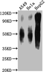
Western Blot
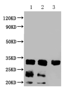
Positive WB detected in: A549 whole cell lysate, Hela whole cell lysate, HepG2 whole cell lysate
All lanes CD63 antibody at 1:1000
Secondary
Goat polyclonal to mouse IgG at 1/50000 dilution
Predicted band size: 30-120 KD KDa
Observed band size: 30-120 KD KDa
Exposure time:1min
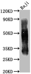
Western Blot
Positive WB detected in: Raji whole cell lysate
All lanes CD63 antibody at 1:1000
Secondary
Goat polyclonal to mouse IgG at 1/50000 dilution
Predicted band size: 30-120 KD KDa
Observed band size: 30-120 KD KDa
Exposure time:1min
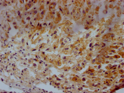
IHC image of CSB-MA004950A0m diluted at 1:500 and staining in paraffin-embedded human glioma tissue performed on a Leica BondTM system. After dewaxing and hydration, antigen retrieval was mediated by high pressure in a citrate buffer (pH 6.0). Section was blocked with 10% normal goat serum 30min at 37°C. Then primary antibody (1% BSA) was incubated at 4°C overnight. The primary is detected by a Goat anti-rabbit IgG labeled by HRP and visualized using 0.05% DAB.
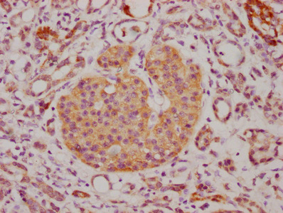
IHC image of CSB-MA004950A0m diluted at 1:500 and staining in paraffin-embedded human lung cancer tissue performed on a Leica BondTM system. After dewaxing and hydration, antigen retrieval was mediated by high pressure in a citrate buffer (pH 6.0). Section was blocked with 10% normal goat serum 30min at 37°C. Then primary antibody (1% BSA) was incubated at 4°C overnight. The primary is detected by a Goat anti-rabbit IgG labeled by HRP and visualized using 0.05% DAB.
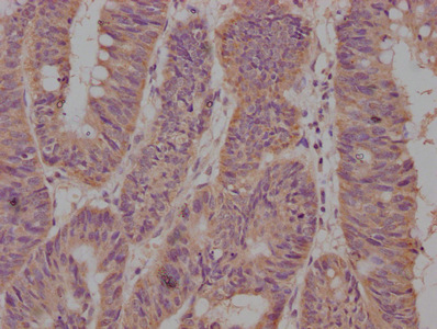
IHC image of CSB-MA004950A0m diluted at 1:500 and staining in paraffin-embedded human lung cancer tissue performed on a Leica BondTM system. After dewaxing and hydration, antigen retrieval was mediated by high pressure in a citrate buffer (pH 6.0). Section was blocked with 10% normal goat serum 30min at 37°C. Then primary antibody (1% BSA) was incubated at 4°C overnight. The primary is detected by a Goat anti-rabbit IgG labeled by HRP and visualized using 0.05% DAB.
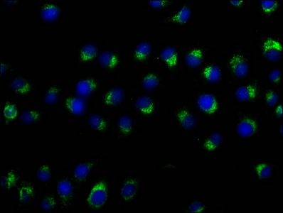
Immunofluorescence staining of A549 cells with CSB-MA004950A0m at 1:50, counter-stained with DAPI. The cells were fixed in 4% formaldehyde and blocked in 10% normal Goat Serum. The cells were then incubated with the antibody overnight at 4°C. Nuclear DNA was labeled in blue with DAPI. The secondary antibody was FITC-conjugated AffiniPure Goat Anti-Mouse IgG (H+L).
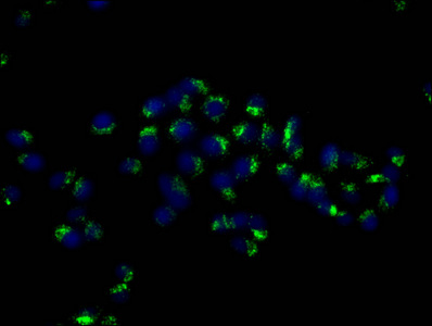
Immunofluorescence staining of Hela cells with CSB-MA004950A0m at 1:50, counter-stained with DAPI. The cells were fixed in 4% formaldehyde and blocked in 10% normal Goat Serum. The cells were then incubated with the antibody overnight at 4°C. Nuclear DNA was labeled in blue with DAPI. The secondary antibody was FITC-conjugated AffiniPure Goat Anti-Mouse IgG (H+L).
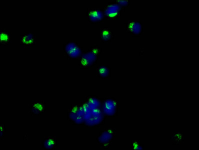
Immunofluorescence staining of MCF-7 cells with CSB-MA004950A0m at 1:50, counter-stained with DAPI. The cells were fixed in 4% formaldehyde and blocked in 10% normal Goat Serum. The cells were then incubated with the antibody overnight at 4°C. Nuclear DNA was labeled in blue with DAPI. The secondary antibody was FITC-conjugated AffiniPure Goat Anti-Mouse IgG (H+L).
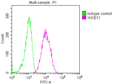
Overlay histogram showing A549 cells stained with CSB-MA004950A0m (red line) at 1:100. The cells were fixed in 4% formaldehyde and permeated by 0.2% TritonX-100. Then 10% normal goat serum was Incubated to block non-specific protein-protein interactions followed by the antibody (1µg/1*106cells) for 1 h at 4°C. The secondary antibody used was FITC-conjugated Goat Anti-Mouse IgG(H+L) at 1/100 dilution for 30min at 4°C. Isotype control antibody (green line) was mouse IgG2b (1µg/1*106cells) used under the same conditions. Acquisition of >10,000 events was performed.
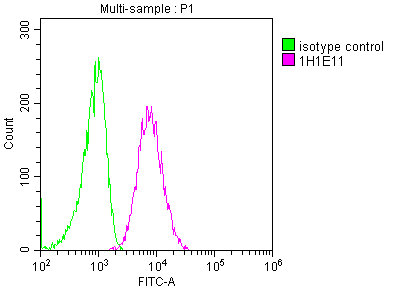
Overlay histogram showing Hela cells stained with CSB-MA004950A0m (red line) at 1:100. The cells were fixed in 4% formaldehyde and permeated by 0.2% TritonX-100. Then 10% normal goat serum was Incubated to block non-specific protein-protein interactions followed by the antibody (1µg/1*106cells) for 1 h at 4°C. The secondary antibody used was FITC-conjugated Goat Anti-Mouse IgG(H+L) at 1/100 dilution for 30min at 4°C. Isotype control antibody (green line) was mouse IgG2b (1µg/1*106cells) used under the same conditions. Acquisition of >10,000 events was performed.
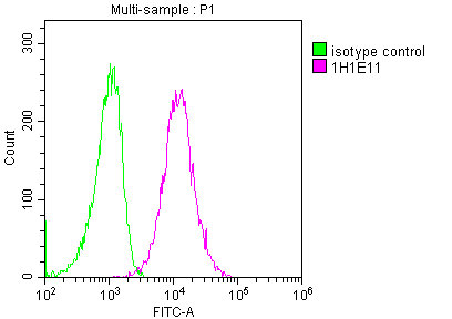
Overlay histogram showing HepG2 cells stained with CSB-MA004950A0m (red line) at 1:100. The cells were fixed in 4% formaldehyde and permeated by 0.2% TritonX-100. Then 10% normal goat serum was Incubated to block non-specific protein-protein interactions followed by the antibody (1µg/1*106cells) for 1 h at 4°C. The secondary antibody used was FITC-conjugated Goat Anti-Mouse IgG(H+L) at 1/100 dilution for 30min at 4°C. Isotype control antibody (green line) was mouse IgG2b (1µg/1*106cells) used under the same conditions. Acquisition of >10,000 events was performed.
CD63 monoclonal antibody is an IgG2a isotype that specifically recognizes CD63 protein. It has been extensively characterized and shown to be highly specific in human samples. This CD63 monoclonal antibody is suitable for use in a variety of research applications, including ELISA, WB, IHC, IF, and FC. It was generated using hybridoma technology by fusing mouse spleen cells immunized with recombinant human CD63 protein (103-203aa) with myeloma cells. The resulting hybridoma clones were screened for those that produce high-affinity CD63 antibodies. The selected hybridomas are cultured in the mouse abdominal cavity, and CD63 monoclonal antibodies are then collected from the mouse ascites.
CD63, also known as LAMP-3, is primarily found in the membranes of late endosomes and lysosomes and is involved in various cellular processes such as antigen presentation, phagocytosis, and intracellular trafficking. CD63 interacts with other tetraspanins, integrins, and other proteins to form tetraspanin-enriched microdomains (TEMs) in the membrane. These TEMs play a role in signal transduction, protein trafficking, and cell adhesion.


Email: support@cusabio.com
Distributors Worldwide



