Tag-Free
The tag type will be determined during production process. If you have specified tag type, please tell us and we will develop the specified tag preferentially.
Buffer
35% FBS + 55% DMEM + 10% DMSO
Storage
Frozen in liquid nitrogen or -80°C
Lead Time
Basically, we can dispatch the products out in 1-3 working days after receiving your orders. Delivery time maybe differs from different purchasing way or location, please kindly consult your local distributors for specific delivery time.
Note: All of our proteins are default shipped with normal blue ice packs, if you request to ship with dry ice, please communicate with us in advance and extra fees will be charged.
Images
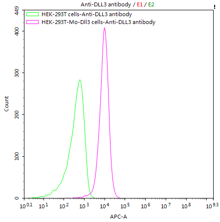
Untransfected HEK-293T cells (green line) and transfected Mouse Dll3 HEK-293T Stable cells (red line) were stained with anti-DLL3 antibody (CSB-RA882142A1HU) (2µg/1*106cells), washed and then followed by APC-conjugated anti-Human IgG Fc antibody and analyzed with flow cytometry.
Inhibits primary neurogenesis. May be required to divert neurons along a specific differentiation pathway. Plays a role in the formation of somite boundaries during segmentation of the paraxial mesoderm.
Gene References into Functions
Structural deformities of the vertebral column and adjacent ribs in the pudgy mouse are caused by mutations in Dll3. Review.PMID:27655002
Dll3 overexpression promoted PI3K/Akt signaling through inhibiting Notch signaling in lung cancer.PMID:28007595
O-fucosylation of DLL3 is required for its function during somitogenesis.PMID:25856312
Intriguing changes are observed in the cranio-caudal borders of multifidus muscle in mutant Dll3 and Lfng models of idiopathic scoliosis.PMID:22095884
Dll3 has a unique function during T-cell development that is distinct from the role played by the other DSL ligands of Notch.PMID:21151194
Dll3 targets Notch1 for lysosomal degradation preventing Notch1 from undergoing post-translational processing.PMID:21147753
Axial skeletal defects caused by mutation in the spondylocostal dysplasia/pudgy gene Dll3 are associated with disruption of the segmentation clock within the presomitic mesoderm.PMID:11923214
DLL3 knockout mice have segmentation and neural defectsPMID:12141422
spondylocostal dysostosis (SCD) is caused by mutation in Delta-like 3 (DLL3), Mesoderm posterior 2 (MESP2), and Lunatic fringe (LFNG); three genes that are components of the Notch signaling pathway.PMID:17600782
study reports that another Notch ligand, Dll3, is expressed in developing hair cells, in a pattern that overlaps that of Dll1 and Jag2; Dll3 may play a role in lateral inhibition similar to that of Dll1 and Jag2PMID:17823936
study shows that genetic interactions between Notch1 and Dll3 result in vertebral segmental defects similar to those seen in congenital scoliosis; craniofacial anomalies not previously observed in Dll3 homozygous animals were identifiedPMID:17849441
Dll3 are expressed in the developing mouse eye and in retinal progenitor cell.PMID:19191219
a complex interplay of E-box binding proteins spatially and temporally regulate Dll3 levels during neural tube development.PMID:19389376
Data show that the ubiquitin ligase Huwe1 operates upstream of the N-Myc-DLL3-Notch pathway to control neural stem cell activity and promote neurogenesis.PMID:19686682
Show
More
Hide
All
Subcellular Location
Membrane; Single-pass type I membrane protein.
Tissue Specificity
Predominantly expressed in the neuroectoderm and paraxial mesoderm during embryogenesis.