Call us
301-363-4651 (Available 9 a.m. to 5 p.m. CST from Monday to Friday)
| Application | Recommended Dilution |
|---|---|
| WB | 1:500-1:5000 |
| IHC | 1:200-1:500 |
| IF | 1:50-1:200 |
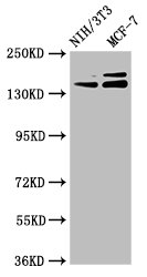
Western Blot
Positive WB detected in: NIH/3T3 whole cell lysate, MCF-7 whole cell lysate
All lanes: UMODL1 antibody at 5.1µg/ml
Secondary
Goat polyclonal to rabbit IgG at 1/50000 dilution
Predicted band size: 145, 157, 137, 150 kDa
Observed band size: 145, 157 kDa
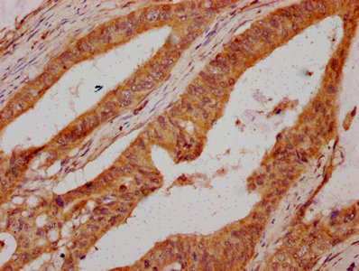
IHC image of CSB-PA025617LA01HU diluted at 1:300 and staining in paraffin-embedded human colon cancer performed on a Leica BondTM system. After dewaxing and hydration, antigen retrieval was mediated by high pressure in a citrate buffer (pH 6.0). Section was blocked with 10% normal goat serum 30min at RT. Then primary antibody (1% BSA) was incubated at 4°C overnight. The primary is detected by a biotinylated secondary antibody and visualized using an HRP conjugated SP system.
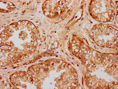
IHC image of CSB-PA025617LA01HU diluted at 1:300 and staining in paraffin-embedded human prostate cancer performed on a Leica BondTM system. After dewaxing and hydration, antigen retrieval was mediated by high pressure in a citrate buffer (pH 6.0). Section was blocked with 10% normal goat serum 30min at RT. Then primary antibody (1% BSA) was incubated at 4°C overnight. The primary is detected by a biotinylated secondary antibody and visualized using an HRP conjugated SP system.
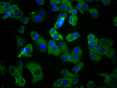
Immunofluorescence staining of MCF-7 cells with CSB-PA025617LA01HU at 1:100, counter-stained with DAPI. The cells were fixed in 4% formaldehyde, permeabilized using 0.2% Triton X-100 and blocked in 10% normal Goat Serum. The cells were then incubated with the antibody overnight at 4°C. The secondary antibody was Alexa Fluor 488-congugated AffiniPure Goat Anti-Rabbit IgG(H+L).
This antibody reacts against the human and mouse UMOLD1 protein. It was raised in rabbit using the partial human UMOLD1 protein as the immunogen. The immunogen region corresponds to the 537-654aa of Recombinant Human UMOLD1 that is a section of the second EGF-like domain. This antibody is a non-conjugate polyclonal IgG with a purity higher than 95% purified by protein G. The uses of this antibody were tested on ELISA, Western blot (WB), Immunohistochemistry (IHC), and Immunofluorescence (IF) applications. The target protein UMOLD1 participates in the response of gonadotropin-releasing hormone, regulates gene expression, and with that, the adipose tissue and ovarian follicle development. The domains of the protein are suggested to bind calcium, interact with proteins, carbohydrate side chains, and other types of substrates. Also, UMOLD1 could have an antiproteinase function, and the presence of the zona pellucida domain (ZP) suggests that the UMOLD1 could have fundamental roles in development, hearing, immunity, or cancer. Indeed, some evidence suggests that it plays a role in high myopia susceptibility.


Email: support@cusabio.com
Distributors Worldwide