It differs from different batches. Please contact us to confirm it.
Buffer
-20°C, pH7.4 PBS, 0.05% NaN3, 40% Glycerol
Tested Applications
ELISA,IHC
Storage
Upon receipt, store at -20°C or -80°C. Avoid repeated freeze.
Lead Time
Basically, we can dispatch the products out in 1-3 working days after receiving your orders. Delivery time maybe differs from different purchasing way or location, please kindly consult your local distributors for specific delivery time.
Note: All of our proteins are default shipped with normal blue ice packs, if you request to ship with dry ice, please communicate with us in advance and extra fees will be charged.
Usage
For Research Use Only. Not for use in diagnostic or therapeutic procedures.
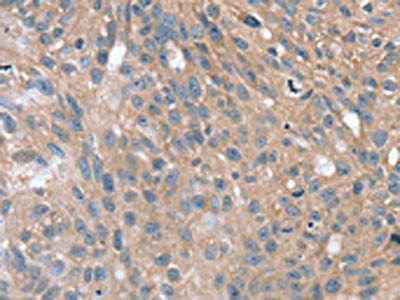
The image on the left is immunohistochemistry of paraffin-embedded Human breast cancer tissue using CSB-PA974733(SPON1 Antibody) at dilution 1/25, on the right is treated with synthetic peptide. (Original magnification: ×200)
Cell adhesion protein that promotes the attachment of spinal cord and sensory neuron cells and the outgrowth of neurites in vitro. May contribute to the growth and guidance of axons in both the spinal cord and the PNS. Major factor for vascular smooth muscle cell.
Gene References into Functions
gene expression experiment revealed that CYP2B6, SPON1, and GSG1L can be activated concomitantly through a constitutive androstane receptor (CAR) activation pathwayPMID:27010727
SPON1 promotes metastatic progression through Fak and Src dependent pathway in human osteosarcoma.PMID:26032498
These data suggest that SPON1 may be associated with the differential rate of cognitive decline in Alzheimer disease.PMID:23535033
High SPON1 expression is associated with high-grade gliomas.PMID:24158112
Genome-wide scan of healthy human connectome discovers SPON1 gene variant influencing dementia severityPMID:23471985
downregulates osteoclast recruitment to the root side of periodontal tissue via low-density lipoprotein receptor-related protein 8 and inhibits differentiation of osteoclastic precursorsPMID:21488757
These findings indicate that F-spondin regulates the differentiation of human cementoblast-like cells via BMP7 expression.PMID:22244873
The unique feature of F-spondin FS domain is the presence of three disulfide bonds associated with the N- and C-termini of the domain and a highly conserved N-linked glycosylation site.PMID:21569239
expression profile of VSGP/F-spondin identifies this molecule as a potential diagnostic marker or target for developing therapeutic strategies to treat ovarian carcinomaPMID:16103746
identified F-spondin as a promoting factor for cementoblastic differentiationPMID:16965763
The crystal structure of the reelin-N domain resolved to 2.0 A reveals a variant immunoglobulin-like fold and potential heparin-binding sites.PMID:18602404
These studies identify F-spondin as a novel protein in osteoarthritis cartilage, where it may act in situ at lesional areas to activate latent TGF-beta and induce cartilage degradation.PMID:18780763
Highest expression in lung, lower expression in brain, heart, kidney, liver and testis, and lowest expression in pancreas, skeletal muscle and ovary. Not expressed in spleen.