
Call us
301-363-4651 (Available 9 a.m. to 5 p.m. CST from Monday to Friday)

| Application | Recommended Dilution |
|---|---|
| WB | 1:500-1:5000 |
| IF | 1:50-1:200 |
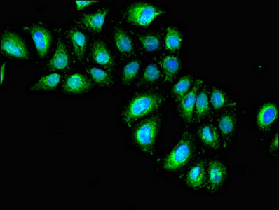
Immunofluorescent analysis of A549 cells using CSB-PA011621EA01HU at dilution of 1: 100 and Alexa Fluor 488-congugated AffiniPure Goat Anti-Rabbit IgG(H+L)
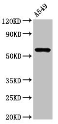
Western Blot
Positive WB detected in: A549 whole cell lysate
All lanes: IL1R1 antibody at 3μg/ml
Secondary
Goat polyclonal to rabbit IgG at 1/50000 dilution
Predicted band size: 66 kDa
Observed band size: 66 kDa
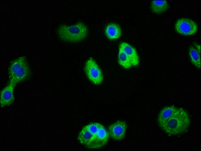
Immunofluorescence staining of HepG2 cells with CSB-PA011621EA01HU at 1:100, counter-stained with DAPI. The cells were fixed in 4% formaldehyde, permeabilized using 0.2% Triton X-100 and blocked in 10% normal Goat Serum. The cells were then incubated with the antibody overnight at 4°C. The secondary antibody was Alexa Fluor 488-congugated AffiniPure Goat Anti-Rabbit IgG(H+L).
IL1R1 polyclonal antibody is synthesized by inoculating rabbits with a recombinant protein corresponding to the amino acids 18-320 of human IL1R1 protein. This IL1R1 antibody is purified from serum using protein G affinity chromatography. Its purity is greater than 95%. It is supplied as a liquid solution in a buffer containing stabilizers. The specificity and sensitivity of this IL1R1 polyclonal antibody for human IL1R1 protein have been verified in ELISA, WB, and IF applications.
IL1R1 protein mainly functions to mediate the biological effects of the pro-inflammatory cytokines IL-1α and IL-1β by activating downstream signaling pathways. IL1R1 is involved in the regulation of the immune response, inflammation, metabolism, and glucose homeostasis, and has been implicated in the development of autoimmune and inflammatory diseases.


Email: support@cusabio.com
Distributors Worldwide



