
Call us
301-363-4651 (Available 9 a.m. to 5 p.m. CST from Monday to Friday)

| Application | Recommended Dilution |
|---|---|
| WB | 1:500-1:5000 |
| IHC | 1:20-1:200 |
| IF | 1:50-1:200 |
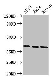
Western Blot
Positive WB detected in: A549 whole cell lysate, Hela whole cell lysate, Mouse brain tissue
All lanes: HLA-C antibody at 3.4µg/ml
Secondary
Goat polyclonal to rabbit IgG at 1/50000 dilution
Predicted band size: 41 kDa
Observed band size: 41 kDa
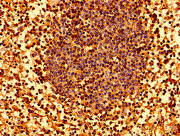
Immunohistochemistry of paraffin-embedded human lymph node tissue using CSB-PA320739LA01HU at dilution of 1:100
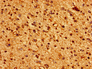
Immunohistochemistry of paraffin-embedded human glioma using CSB-PA320739LA01HU at dilution of 1:100
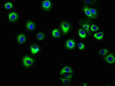
Immunofluorescence staining of HepG2 cells with CSB-PA320739LA01HU at 1:166, counter-stained with DAPI. The cells were fixed in 4% formaldehyde, permeabilized using 0.2% Triton X-100 and blocked in 10% normal Goat Serum. The cells were then incubated with the antibody overnight at 4°C. The secondary antibody was Alexa Fluor 488-congugated AffiniPure Goat Anti-Rabbit IgG(H+L).
The HLA-C antibody can be used to recognize the human and mouse HLA-C protein. A rabbit got immunization with the recombinant human HLA class I histocompatibility antigen, Cw-7 alpha chain protein (62-306aa). After that, an immune response occurred in the rabbit producing large amounts of antibodies against the HLA-C protein. The rabbit's blood was collected and isolated to get the anti-serum, which was protein G-purified to obtain the HLA-C polyclonal antibody (purity 95%+). This HLA-C antibody has accessed its specificity in ELISA, WB, IHC, and IF applications.
The HLA class I histocompatibility antigen, Cw-7 alpha chain protein mainly functions to present peptides derived from intracellular proteins to CD8+ T cells for immune recognition, helping to regulate the immune response and protect the body against infected or abnormal cells.


Email: support@cusabio.com
Distributors Worldwide



