
Call us
301-363-4651 (Available 9 a.m. to 5 p.m. CST from Monday to Friday)

| Application | Recommended Dilution |
|---|---|
| IHC | 1:20-1:200 |
| IF | 1:20-1:200 |
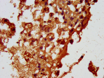
IHC image of CSB-PA857429LA01HU diluted at 1:100 and staining in paraffin-embedded human melanoma performed on a Leica BondTM system. After dewaxing and hydration, antigen retrieval was mediated by high pressure in a citrate buffer (pH 6.0). Section was blocked with 10% normal goat serum 30min at RT. Then primary antibody (1% BSA) was incubated at 4°C overnight. The primary is detected by a biotinylated secondary antibody and visualized using an HRP conjugated SP system.
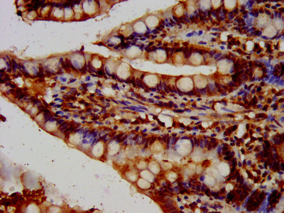
IHC image of CSB-PA857429LA01HU diluted at 1:100 and staining in paraffin-embedded human small intestine tissue performed on a Leica BondTM system. After dewaxing and hydration, antigen retrieval was mediated by high pressure in a citrate buffer (pH 6.0). Section was blocked with 10% normal goat serum 30min at RT. Then primary antibody (1% BSA) was incubated at 4°C overnight. The primary is detected by a biotinylated secondary antibody and visualized using an HRP conjugated SP system.
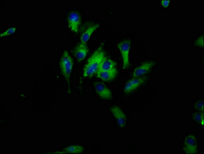
Immunofluorescence staining of Hela cells with CSB-PA857429LA01HU at 1:33, counter-stained with DAPI. The cells were fixed in 4% formaldehyde, permeabilized using 0.2% Triton X-100 and blocked in 10% normal Goat Serum. The cells were then incubated with the antibody overnight at 4°C. The secondary antibody was Alexa Fluor 488-congugated AffiniPure Goat Anti-Rabbit IgG(H+L).
The HBEGF Antibody is produced by immunizing rabbits with the peptide mapping within amino acids 63-148 of the human HBEGF protein. This polyclonal HBEGF antibody exists as an unconjugated IgG. It is purified by protein G and reaches up to 95% in purity. It only reacts with human HBEGF protein. Quality tests of this antibody in ELISA, IHC, and IF have been verified. Interactions between HBEGF and EGF receptors are involved in multiple biological processes, including wounding healing, blast implantation, and tumorigenesis.


Email: support@cusabio.com
Distributors Worldwide



