GPR34 Antibody

Code: CSB-PA891571LA01HU
Size:
20μg
20μg50μg100μg
US$83
Quantity:
Species Reactivity: Human, Mouse
Raised in: Rabbit
Application: ELISA, WB, IHC, IF
For inquiries on large quantities or another requirements
Send an Inquiry
Start an on-line Chat
Online ordering is currently available for U.S. customers only. For orders outside the U.S., please kindly submit an inquiry or start a chat with us.

Product Details

Uniprot NO.
Target Names
GPR34
Alternative Names
GPR34; Probable G-protein coupled receptor 34
Raised in
Rabbit
Species Reactivity
Human, Mouse
Immunogen
Recombinant Human Probable G-protein coupled receptor 34 protein (1-61AA)
Immunogen Species
Homo sapiens (Human)
Conjugate
Non-conjugated
Isotype
IgG
Purification Method
>95%, Protein G purified
Concentration
It differs from different batches. Please contact us to confirm it.
Buffer
Preservative: 0.03% Proclin 300
Constituents: 50% Glycerol, 0.01M PBS, PH 7.4
Tested Applications
ELISA, WB, IHC, IF
Recommended Dilution
ApplicationRecommended Dilution
WB1:500-1:2000
IHC1:50-1:200
IF1:20-1:100
Storage
Upon receipt, store at -20°C or -80°C. Avoid repeated freeze.
Lead Time
Basically, we can dispatch the products out in 1-3 working days after receiving your orders. Delivery time maybe differs from different purchasing way or location, please kindly consult your local distributors for specific delivery time.
Note: All of our proteins are default shipped with normal blue ice packs, if you request to ship with dry ice, please communicate with us in advance and extra fees will be charged.
Usage
For Research Use Only. Not for use in diagnostic or therapeutic procedures.
Datasheet & COA
Images
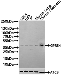
  • Western Blot
    Positive WB detected in: U251 whole cell lysate(30µg), U87 whole cell lysate(30µg), JK whole cell lysate(30µg), Mouse Lung tissue lysate(30µg), Mouse Stomach tissue lysate(30µg)
    All lanes: GPR34 antibody at 1:1000
    Secondary
    Goat polyclonal to rabbit IgG at 1/20000 dilution
    Predicted band size: 44 kDa
    Observed band size: 44 kDa
    Exposure time: 120s
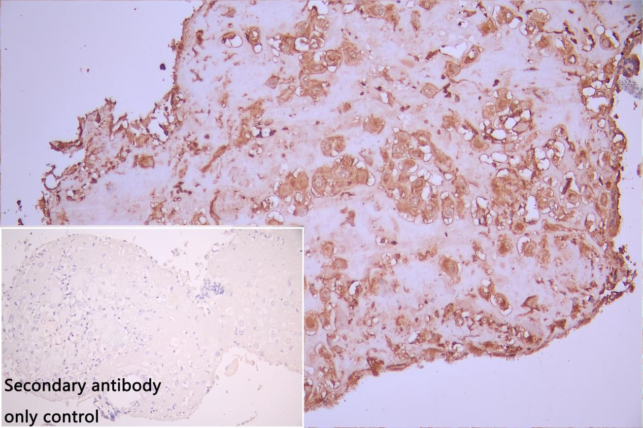
  • IHC image of CSB-PA891571LA01HU diluted at 1:50 and staining in paraffin-embedded human placenta tissue performed on a Leica BondTM system. After dewaxing and hydration, antigen retrieval was mediated by high pressure in a citrate buffer (pH 6.0). Section was blocked with 10% normal goat serum 30min at RT. Then primary antibody (1% BSA) was incubated at 4°C overnight. The primary is detected by a Goat anti-rabbit polymer IgG labeled by HRP and visualized using 0.05% DAB. Secondary antibody only control: uses 1% BSA instead of primary antibody
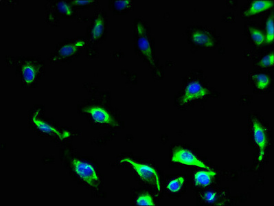
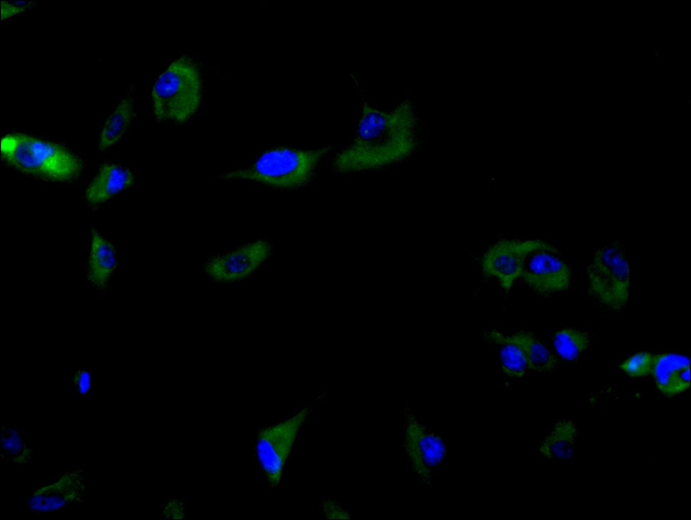
  • Immunofluorescence staining of U251 cell with CSB-PA891571LA01HU at 1:30, counter-stained with DAPI. The cells were fixed in 4% formaldehyde and blocked in 10% normal Goat Serum. The cells were then incubated with the antibody overnight at 4C. The secondary antibody was Alexa Fluor 488-congugated AffiniPure Goat Anti-Rabbit IgG(H+L).
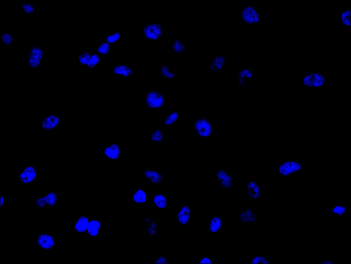
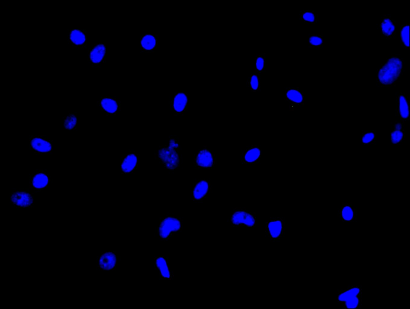
  • Immunofluorescence staining of U251 cell with 5% goat serum, counter-stained with DAPI. The cells were fixed in 4% formaldehyde and blocked in 10% normal Goat Serum. The cells were then incubated with the antibody overnight at 4C. The secondary antibody was Alexa Fluor 488-congugated AffiniPure Goat Anti-Rabbit IgG(H+L).
  • Immunofluorescence staining of U87 cell with CSB-PA891571LA01HU at 1:30, counter-stained with DAPI. The cells were fixed in 4% formaldehyde and blocked in 10% normal Goat Serum. The cells were then incubated with the antibody overnight at 4C. The secondary antibody was Alexa Fluor 488-congugated AffiniPure Goat Anti-Rabbit IgG(H+L).
  • Immunofluorescence staining of U87 cell with 5% goat serum, counter-stained with DAPI. The cells were fixed in 4% formaldehyde and blocked in 10% normal Goat Serum. The cells were then incubated with the antibody overnight at 4C. The secondary antibody was Alexa Fluor 488-congugated AffiniPure Goat Anti-Rabbit IgG(H+L).

Customer Reviews and Q&A

 Customer Reviews

Target Background

Function(From Uniprot)
Orphan receptor.
Gene References into Functions
  1. By monitoring fused FLAG-tag and conformation-sensitive native epitope during expression of GPR34 mutants, a tri-basic motif in the first intracellular loop was identified as key topogenic signal that dictates the orientation of transmembrane domain-1. PMID:27086875
  2. GPR34 knockdown impairs the proliferation and migration of HGC-27 gastric cancer cells in vitro and provides a potential implication for therapy of gastric cancer. PMID:25673461
  3. up-regulation of GPR34 expression in human gastric carcinoma may play a critical role in tumor progression and in determining patient prognosis PMID:23836308
  4. The present studies confirm that GPR34 is a cellular receptor for LysoPS, especially with a fatty acid at the sn-2 position. PMID:22343749
  5. these results are the first to identify a role for a GPR34 in lymphoma cell growth, provide insight into GPR34-mediated signaling, identify a genetically unique subset of MZLs that express high levels of GPR34. PMID:22966169
  6. data show that multiple translation initiation starts and alternative splicing contribute to the supragenomic diversification of GPR34 PMID:16338117

Show More

Hide All

Subcellular Location
Cell membrane; Multi-pass membrane protein.
Protein Families
G-protein coupled receptor 1 family
Tissue Specificity
Broadly expressed.
Database Links

HGNC: 4490

UNIGENE: Hs.495989

KEGG: hsa:2857

STRING: 9606.ENSP00000367378

OMIM: 300241

icon of phone
Call us
301-363-4651 (Available 9 a.m. to 5 p.m. CST from Monday to Friday)

icon of address
Address
No. 269, Shendun 5th Road, Donghu Hi-Tech Development Area, Hubei Province, 430206, P.R.China
icon of social media
Join us with

Subscribe newsletter

Leave a message

* To protect against spam, please pass the CAPTCHA test below.
CAPTCHA verification
© 2007-2025 CUSABIO TECHNOLOGY LLC All rights reserved. 鄂ICP备15011166号-1
Select 0 Products