
Call us
301-363-4651 (Available 9 a.m. to 5 p.m. CST from Monday to Friday)

| Application | Recommended Dilution |
|---|---|
| WB | 1:1000-1:5000 |
| IHC | 1:1000-1:2000 |
| IF | 1:200-1:500 |
| IP | 1:200-1:2000 |
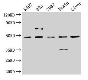
Western Blot
Positive WB detected in: K562 whole cell lysate, 293 whole cell lysate, 293T whole cell lysate, Rat brain tissue, Mouse liver tissue
All lanes: CSNK1D antibody at 5µg/ml
Secondary
Goat polyclonal to rabbit IgG at 1/50000 dilution
Predicted band size: 48, 47 kDa
Observed band size: 48 kDa
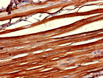
IHC image of CSB-PA006067LA01HU diluted at 1:1000 and staining in paraffin-embedded human skeletal muscle tissue performed on a Leica BondTM system. After dewaxing and hydration, antigen retrieval was mediated by high pressure in a citrate buffer (pH 6.0). Section was blocked with 10% normal goat serum 30min at RT. Then primary antibody (1% BSA) was incubated at 4°C overnight. The primary is detected by a biotinylated secondary antibody and visualized using an HRP conjugated SP system.
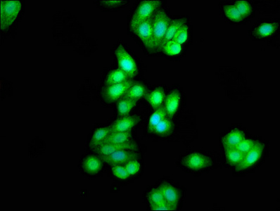
Immunofluorescence staining of HepG2 cells with CSB-PA006067LA01HU at 1:333, counter-stained with DAPI. The cells were fixed in 4% formaldehyde, permeabilized using 0.2% Triton X-100 and blocked in 10% normal Goat Serum. The cells were then incubated with the antibody overnight at 4°C. The secondary antibody was Alexa Fluor 488-congugated AffiniPure Goat Anti-Rabbit IgG(H+L).
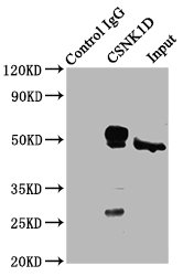
Immunoprecipitating CSNK1D in Hela whole cell lysate
Lane 1: Rabbit control IgG (1µg) instead of CSB-PA006067LA01HU in Hela whole cell lysate. For western blotting, a HRP-conjugated Protein G antibody was used as the secondary antibody (1/2000)
Lane 2: CSB-PA006067LA01HU (6µg) + Hela whole cell lysate (500µg)
Lane 3: Hela whole cell lysate (10µg)
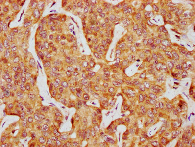
IHC image of CSB-PA006067LA01HU diluted at 1:1000 and staining in paraffin-embedded human liver cancer performed on a Leica BondTM system. After dewaxing and hydration, antigen retrieval was mediated by high pressure in a citrate buffer (pH 6.0). Section was blocked with 10% normal goat serum 30min at RT. Then primary antibody (1% BSA) was incubated at 4°C overnight. The primary is detected by a biotinylated secondary antibody and visualized using an HRP conjugated SP system.
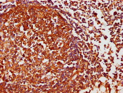
IHC image of CSB-PA006067LA01HU diluted at 1:1000 and staining in paraffin-embedded human lymph node tissue performed on a Leica BondTM system. After dewaxing and hydration, antigen retrieval was mediated by high pressure in a citrate buffer (pH 6.0). Section was blocked with 10% normal goat serum 30min at RT. Then primary antibody (1% BSA) was incubated at 4°C overnight. The primary is detected by a biotinylated secondary antibody and visualized using an HRP conjugated SP system.
The polyclonal antibody targeting CSNK1D was produced in rabbits immunizing with the recombinant human casein kinase I isoform delta protein (152-398aa). The antibody was subsequently isolated and underwent protein G affinity chromatography to achieve a purity level exceeding 95%. Its specificity encompasses the recognition and binding of the CSNK1D protein in human, rat, and mouse samples.
The CSNK1D polyclonal antibody has been validated for use in ELISA, WB, IHC, IF, and IP applications. This antibody can thus be used for qualitative and quantitative evaluation, localization, and isolation of the CSNK1D protein.


Email: support@cusabio.com
Distributors Worldwide



