Call us
301-363-4651 (Available 9 a.m. to 5 p.m. CST from Monday to Friday)
| Application | Recommended Dilution |
|---|---|
| WB | 1:4000-1:256000 |
| IHC | 1:200-1:500 |
| IF | 1:150-1:300 |
| FC | 1:50-1:200 |
| IP | 1µ |
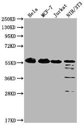
Western Blot
Positive WB detected in: Hela whole cell lysate, MCF-7 whole cell lysate, Jurkat whole cell lysate, NIH/3T3 whole cell lysate
All lanes: PKM antibody at 1:4000
Secondary
Goat polyclonal to Mouse IgG at 1/10000 dilution
Predicted band size: 58 kDa
Observed band size: 58 KDa
Exposure time: 1min
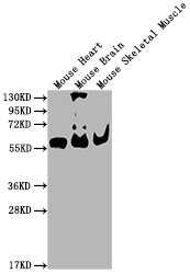
Western Blot
Positive WB detected in: Mouse Heart tissue, Mouse Brain tissue, Mouse Skeletal Muscle tissue
All lanes: PKM antibody at 1:4000
Secondary
Goat polyclonal to Mouse IgG at 1/10000 dilution
Predicted band size: 58 kDa
Observed band size: 58 KDa
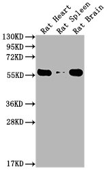
Western Blot
Positive WB detected in: Rat Heart tissue, Rat Spleen tissue, Rat Brain tissue
All lanes: PKM antibody at 1:4000
Secondary
Goat polyclonal to Mouse IgG at 1/10000 dilution
Predicted band size: 55-60 kDa
Observed band size: 55-60 kDa
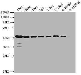
Western Blot
Positive WB detected in: MCF-7 whole cell lysate at 40µg, 20µg, 10µg, 5µg, 2.5µg, 1.25µg, 0.625µg, 0.3125µg
All lanes: PKM antibody at 1:4000
Secondary
Goat polyclonal to Mouse IgG at 1/10000 dilution
Predicted band size: 58 kDa
Observed band size: 58 KDa
Exposure time: 5min
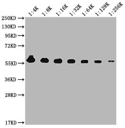
Western Blot
Positive WB detected in: MCF-7 whole cell lysate
All lanes: PKM antibody at 1:4000, 1:8000, 1:16000, 1:32000, 1:64000, 1:128000, 1:256000
Secondary
Goat polyclonal to Mouse IgG at 1/10000 dilution
Predicted band size: 58 kDa
Observed band size: 58 KDa
Exposure time: 5min
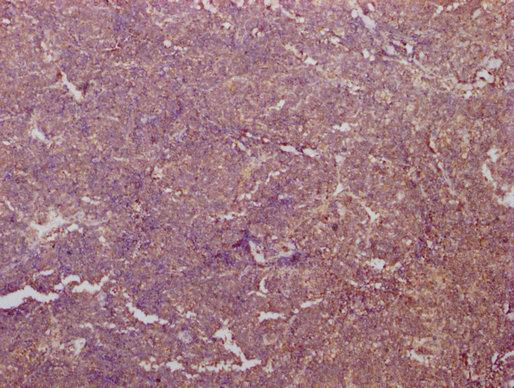
IHC image of CSB-MA018072A0m diluted at 1:400 and staining in paraffin-embedded human tonsil tissue performed on a Leica BondTM system. After dewaxing and hydration, antigen retrieval was mediated by high pressure in a citrate buffer (pH 6.0). Section was blocked with 10% normal goat serum 30min at RT. Then primary antibody (1% BSA) was incubated at 4°C overnight. The primary is detected by a biotinylated secondary antibody and visualized using an HRP conjugated SP system.
IHC image of CSB-MA018072A0m diluted at 1:400 and staining in paraffin-embedded human tonsil tissue performed on a Leica BondTM system. After dewaxing and hydration, antigen retrieval was mediated by high pressure in a citrate buffer (pH 6.0). Section was blocked with 10% normal goat serum 30min at RT. Then primary antibody (1% BSA) was incubated at 4°C overnight. The primary is detected by a biotinylated secondary antibody and visualized using an HRP conjugated SP system.
IHC image of CSB-MA018072A0m diluted at 1:400 and staining in paraffin-embedded human tonsil tissue performed on a Leica BondTM system. After dewaxing and hydration, antigen retrieval was mediated by high pressure in a citrate buffer (pH 6.0). Section was blocked with 10% normal goat serum 30min at RT. Then primary antibody (1% BSA) was incubated at 4°C overnight. The primary is detected by a biotinylated secondary antibody and visualized using an HRP conjugated SP system.
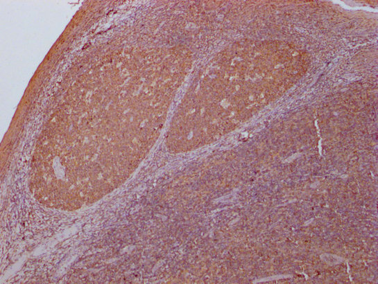
IHC image of CSB-MA018072A0m diluted at 1:400 and staining in paraffin-embedded human lung cancer tissue performed on a Leica BondTM system. After dewaxing and hydration, antigen retrieval was mediated by high pressure in a citrate buffer (pH 6.0). Section was blocked with 10% normal goat serum 30min at RT. Then primary antibody (1% BSA) was incubated at 4°C overnight. The primary is detected by a biotinylated secondary antibody and visualized using an HRP conjugated SP system.
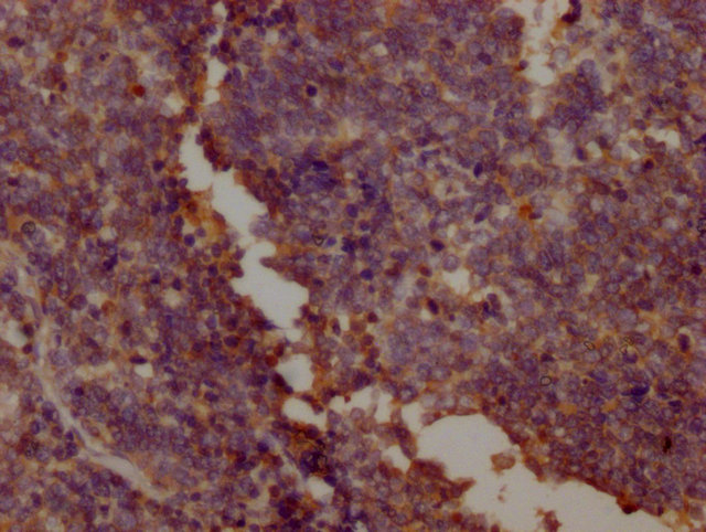
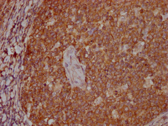
IHC image of CSB-MA018072A0m diluted at 1:400 and staining in paraffin-embedded human lung cancer tissue performed on a Leica BondTM system. After dewaxing and hydration, antigen retrieval was mediated by high pressure in a citrate buffer (pH 6.0). Section was blocked with 10% normal goat serum 30min at RT. Then primary antibody (1% BSA) was incubated at 4°C overnight. The primary is detected by a biotinylated secondary antibody and visualized using an HRP conjugated SP system.
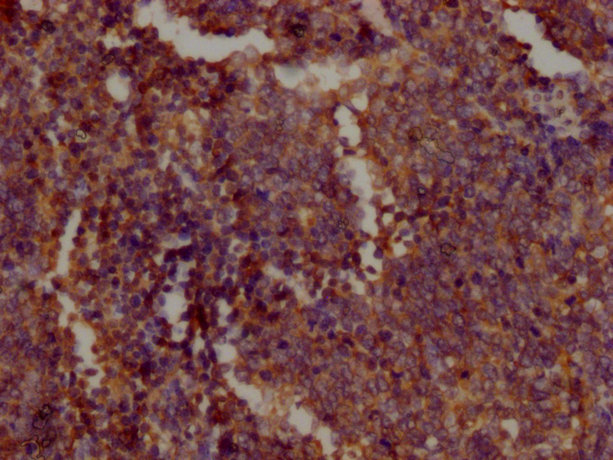
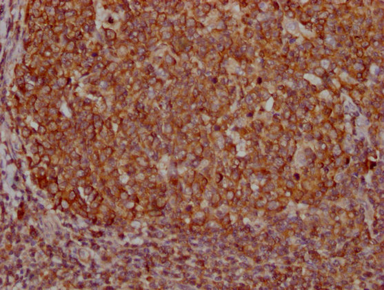
IHC image of CSB-MA018072A0m diluted at 1:400 and staining in paraffin-embedded human lung cancer tissue performed on a Leica BondTM system. After dewaxing and hydration, antigen retrieval was mediated by high pressure in a citrate buffer (pH 6.0). Section was blocked with 10% normal goat serum 30min at RT. Then primary antibody (1% BSA) was incubated at 4°C overnight. The primary is detected by a biotinylated secondary antibody and visualized using an HRP conjugated SP system.
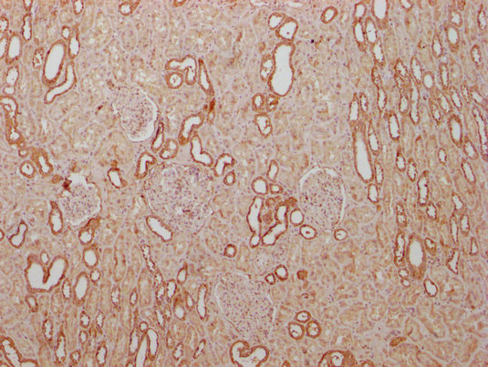
IHC image of CSB-MA018072A0m diluted at 1:400 and staining in paraffin-embedded human kidney tissue performed on a Leica BondTM system. After dewaxing and hydration, antigen retrieval was mediated by high pressure in a citrate buffer (pH 6.0). Section was blocked with 10% normal goat serum 30min at RT. Then primary antibody (1% BSA) was incubated at 4°C overnight. The primary is detected by a biotinylated secondary antibody and visualized using an HRP conjugated SP system.
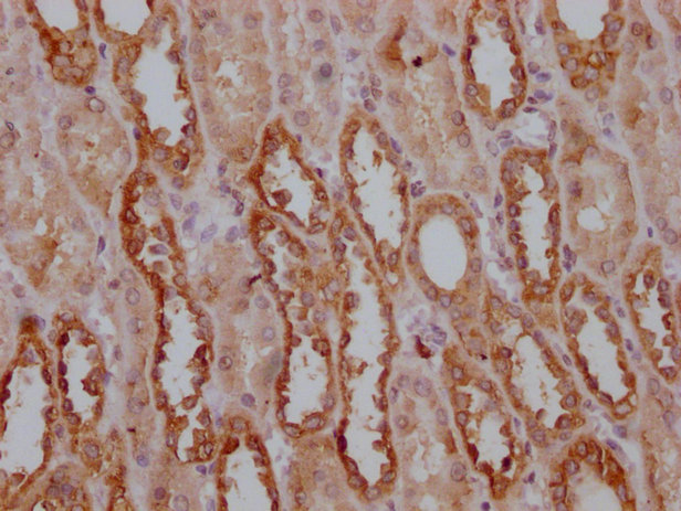
IHC image of CSB-MA018072A0m diluted at 1:400 and staining in paraffin-embedded human kidney tissue performed on a Leica BondTM system. After dewaxing and hydration, antigen retrieval was mediated by high pressure in a citrate buffer (pH 6.0). Section was blocked with 10% normal goat serum 30min at RT. Then primary antibody (1% BSA) was incubated at 4°C overnight. The primary is detected by a biotinylated secondary antibody and visualized using an HRP conjugated SP system.
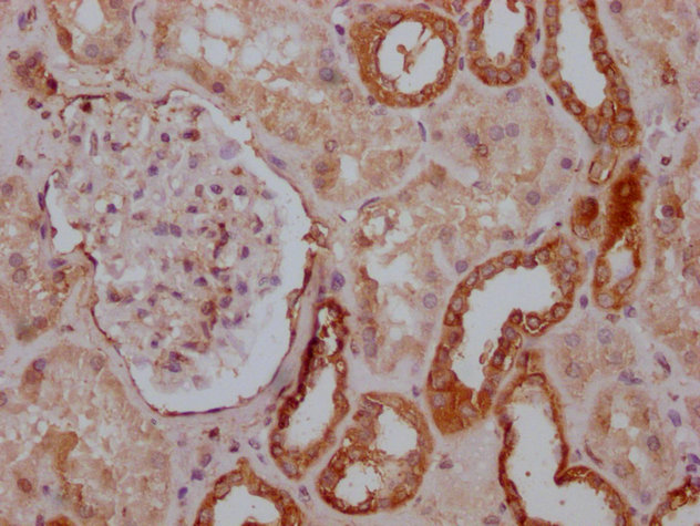
IHC image of CSB-MA018072A0m diluted at 1:400 and staining in paraffin-embedded human kidney tissue performed on a Leica BondTM system. After dewaxing and hydration, antigen retrieval was mediated by high pressure in a citrate buffer (pH 6.0). Section was blocked with 10% normal goat serum 30min at RT. Then primary antibody (1% BSA) was incubated at 4°C overnight. The primary is detected by a biotinylated secondary antibody and visualized using an HRP conjugated SP system.
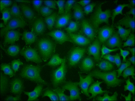
Immunofluorescence staining of A549 cells with CSB-MA018072A0m at 1:230, counter-stained with DAPI. The cells were blocked in 10% normal Goat Serum and then incubated with the primary antibody overnight at 4°C. The secondary antibody was Alexa Fluor 488-congugated AffiniPure Goat Anti-Mouse IgG(H+L).
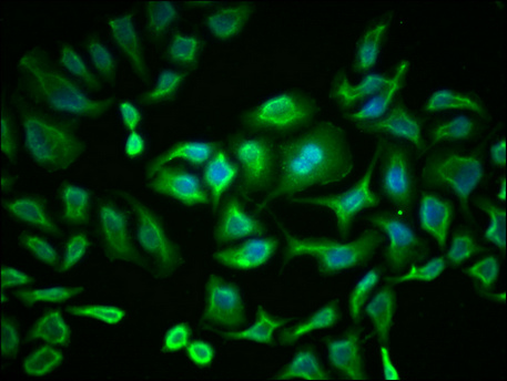
Immunofluorescence staining of Hela cells with CSB-MA018072A0m at 1:230, counter-stained with DAPI. The cells were blocked in 10% normal Goat Serum and then incubated with the primary antibody overnight at 4°C. The secondary antibody was Alexa Fluor 488-congugated AffiniPure Goat Anti-Mouse IgG(H+L).
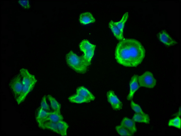
Immunofluorescence staining of HepG2 cells with CSB-MA018072A0m at 1:230, counter-stained with DAPI. The cells were blocked in 10% normal Goat Serum and then incubated with the primary antibody overnight at 4°C. The secondary antibody was Alexa Fluor 488-congugated AffiniPure Goat Anti-Mouse IgG(H+L).
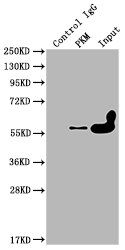
Immunoprecipitating PKM in Hela whole cell lysate
Lane 1: Mouse control IgG instead of CSB-MA018072A0m in Hela whole cell lysate.
Lane 2: CSB-MA018072A0m (1ul) + Hela whole cell lysate (500ug)
Lane 3: Hela whole cell lysate (10ug)
For western blotting, the blot was detected with CSB-MA018072A0m at 1:2000, and a HRP-conjugated Protein G antibody was used as the secondary antibody at 1:2000
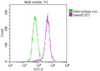
Overlay histogram showing Hela cells stained with CSB-MA018072A0m (red line) at 1:100. The cells were incubated in 1x PBS /10% normal goat serum to block non-specific protein-protein interactions followed by primary antibody for 1 h at 4°C. The secondary antibody used was FITC goat anti-mouse IgG(H+L) at 1/200 dilution for 1 h at 4°C. Isotype control antibody (green line) was used under the same conditions. Acquisition of >10,000 events was performed.
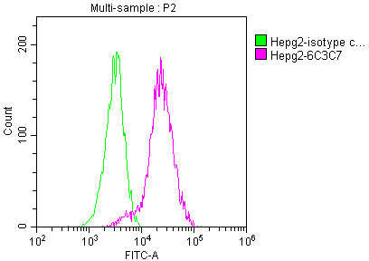
Overlay histogram showing HepG2 cells stained with CSB-MA018072A0m (red line) at 1:100. The cells were incubated in 1x PBS /10% normal goat serum to block non-specific protein-protein interactions followed by primary antibody for 1 h at 4°C. The secondary antibody used was FITC goat anti-mouse IgG(H+L) at 1/200 dilution for 1 h at 4°C. Isotype control antibody (green line) was used under the same conditions. Acquisition of >10,000 events was performed.
CUSABIO has built a mature production system of monoclonal antibodies. Take this PKM monoclonal antibody for example. In the production, the mouses were immunized with Recombinant Human Pyruvate kinase PKM protein at first, and then the spleen cells of the mouse were obtained. Next, used cell hybridization technology to fuse myeloma cells and B lymphocytes in the spleen cells to generate hybridoma. And then screened the obtained hybridoma cells to select the ones that could secrete high-titer antibodies for the production of the PKM monoclonal antibody. This antibody was purified by Protein G and validated in ELISA, WB, IHC, IF, FC, IP.
PKM2 has two isozymes of M type and L type, and M type has M1 and M2 subtypes. M1 is distributed in myocardium, skeletal muscle, and brain tissue; M2 is widely present in tissues during the embryonic period. As the embryo develops, it is gradually replaced by other subtypes in some tissues, and is only highly expressed in cells with a high nucleic acid synthesis rate. , Especially in tumor cells, it is of great significance to the metabolism of cancer and the promotion of tumor formation. At the same time, in the process of tumor progression, due to tumor cell necrosis and metastasis, PKM2 will be released into the blood, and the PKM2 of gastrointestinal tumors It can also be excreted with the stool of cancer patients. Some studies have found that continuous monitoring of PKM2 levels in the plasma of patients with gastric cancer, non-small cell lung cancer, rectal cancer, breast cancer, hepatocellular carcinoma, and renal clear cell carcinoma Comparative analysis shows that the level of PKM2 is significantly increased in tumor patients, which will help the early diagnosis and prognostic judgment of these tumors. Therefore, PKM2 is also used as a very potential new tumor marker. It is expected to become an important anti-tumor drug target.

Email: support@cusabio.com
Distributors Worldwide