Call us
301-363-4651 (Available 9 a.m. to 5 p.m. CST from Monday to Friday)
| Application | Recommended Dilution |
|---|---|
| WB | 1:500-1:10000 |
| IHC | 1:50-1:200 |
| IF | 1:50-1:200 |
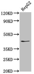
Western Blot
Positive WB detected in: HepG2 whole cell lysate
All lanes: OCT4 antibody at 1:500
Secondary
Goat polyclonal to Mouse IgG at 1/10000 dilution
Predicted band size: 39, 31 kDa
Observed band size: 45 kDa
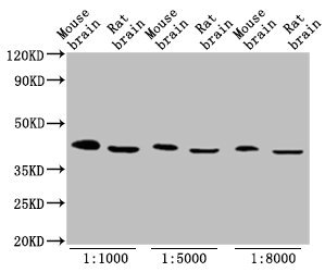
Western Blot
Positive WB detected in: Mouse brain tissue, Rat brain tissue
All lanes: OCT4 antibody at 1:1000, 1:5000, 1:8000
Secondary
Goat polyclonal to Mouse IgG at 1/10000 dilution
Predicted band size: 39, 31 kDa
Observed band size: 45 kDa
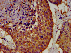
IHC image of CSB-MA018403A0m diluted at 1:100 and staining in paraffin-embedded human lung cancer performed on a Leica BondTM system. After dewaxing and hydration, antigen retrieval was mediated by high pressure in a citrate buffer (pH 6.0). Section was blocked with 10% normal goat serum 30min at RT. Then primary antibody (1% BSA) was incubated at 4°C overnight. The primary is detected by a biotinylated secondary antibody and visualized using an HRP conjugated SP system.
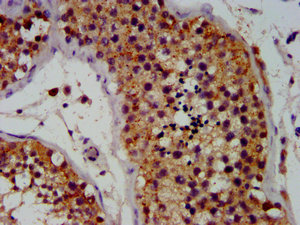
IHC image of CSB-MA018403A0m diluted at 1:100 and staining in paraffin-embedded human testis tissue performed on a Leica BondTM system. After dewaxing and hydration, antigen retrieval was mediated by high pressure in a citrate buffer (pH 6.0). Section was blocked with 10% normal goat serum 30min at RT. Then primary antibody (1% BSA) was incubated at 4°C overnight. The primary is detected by a biotinylated secondary antibody and visualized using an HRP conjugated SP system.
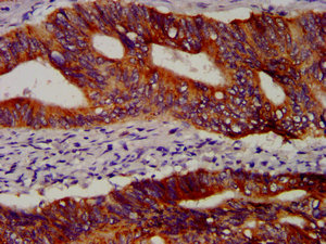
IHC image of CSB-MA018403A0m diluted at 1:100 and staining in paraffin-embedded human colon cancer performed on a Leica BondTM system. After dewaxing and hydration, antigen retrieval was mediated by high pressure in a citrate buffer (pH 6.0). Section was blocked with 10% normal goat serum 30min at RT. Then primary antibody (1% BSA) was incubated at 4°C overnight. The primary is detected by a biotinylated secondary antibody and visualized using an HRP conjugated SP system.
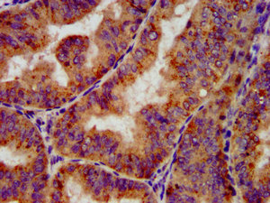
IHC image of CSB-MA018403A0m diluted at 1:100 and staining in paraffin-embedded human endometrial cancer performed on a Leica BondTM system. After dewaxing and hydration, antigen retrieval was mediated by high pressure in a citrate buffer (pH 6.0). Section was blocked with 10% normal goat serum 30min at RT. Then primary antibody (1% BSA) was incubated at 4°C overnight. The primary is detected by a biotinylated secondary antibody and visualized using an HRP conjugated SP system.
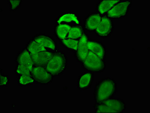
Immunofluorescence staining of A549 cells with CSB-MA018403A0m at 1:100, counter-stained with DAPI. The cells were blocked in 10% normal Goat Serum and then incubated with the primary antibody overnight at 4°C. The secondary antibody was Alexa Fluor 488-congugated AffiniPure Goat Anti-Mouse IgG(H+L).
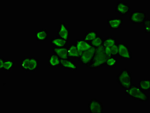
Immunofluorescence staining of Ntera-2 cells with CSB-MA018403A0m at 1:100, counter-stained with DAPI. The cells were blocked in 10% normal Goat Serum and then incubated with the primary antibody overnight at 4°C. The secondary antibody was Alexa Fluor 488-congugated AffiniPure Goat Anti-Mouse IgG(H+L).
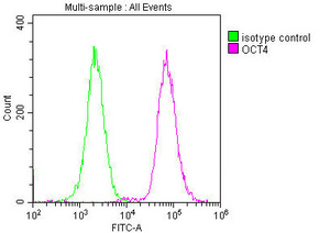
Overlay histogram showing A549 cells stained with CSB-MA018403A0m (red line) at 1:100. The cells were incubated in 1x PBS /10% normal goat serum to block non-specific protein-protein interactions followed by primary antibody for 1 h at 4°C. The secondary antibody used was FITC goat anti-mouse IgG(H+L) at 1/200 dilution for 1 h at 4°C. Isotype control antibody (green line) was used under the same conditions. Acquisition of >10,000 events was performed.
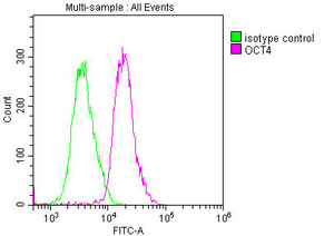
Overlay histogram showing Ntera-2 cells stained with CSB-MA018403A0m (red line) at 1:100. The cells were incubated in 1x PBS /10% normal goat serum to block non-specific protein-protein interactions followed by primary antibody for 1 h at 4°C. The secondary antibody used was FITC goat anti-mouse IgG(H+L) at 1/200 dilution for 1 h at 4°C. Isotype control antibody (green line) was used under the same conditions. Acquisition of >10,000 events was performed.
The OCT4 Monoclonal Antibody is formed by introducing Recombinant Human POU Domain, Class 5, Transcription Factor 1 Protein (1-360AA) in mice. It is an IgG2b subtype antibody that reacts with Human, Mouse, Rat OCT4, a POU transcription factor.
OCT4 is expressed in embryonic stem cells. In these stem cells, it controls the expression of genes that facilitate proliferation. At certain levels, OCT4 alongside Sox2 facilitates the renewal and maintains the pluripotency of stem cells. A fall or rise in OCT4 will activate genes that trigger the differentiation of these germ cells.
Abnormal expression of OCT4 is known to play a role in the embryogenesis and proliferation of cancer cells. The OCT4 Antibody binds to OCT4 in cancer cells. Thus, it can be used for detecting OCT4 and for studying anti-OCT4 options.


Email: support@cusabio.com
Distributors Worldwide