Call us
301-363-4651 (Available 9 a.m. to 5 p.m. CST from Monday to Friday)
| Application | Recommended Dilution |
|---|---|
| WB | 1:10000-1:256000 |
| IHC | 1:100-1:500 |
| FC | 1:100-1:300 |
| IP | 1μl-4μl |
Western Blot
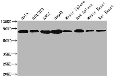
Positive WB detected in: Hela whole cell lysate, NIH/3T3 whole cell lysate, K562 whole cell lysate, HepG2 whole cell lysate, Mouse spleen tissue, Rat spleen tissue, Mouse heart tissue, Rat heart tissue
All lanes HSPA8 antibody at 1:2000
Secondary
Goat polyclonal to mouse IgG at 1/50000 dilution
Predicted band size: 70~75 KDa
Observed band size: 70~75 KDa
Exposure time: 10s
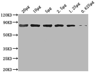
Western Blot
Positive WB detected in: Hela whole cell lysate at 20μg, 10μg, 5μg, 2.5μg, 1.25μg, 0.625μg
All lanes: HSPA8 antibody at 1:2000
Secondary
Goat polyclonal to mouse IgG at 1/50000 dilution
Predicted band size: 70~75 KDa
Observed band size: 70~75 KDa
Exposure time: 10s
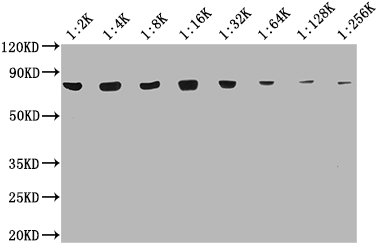
Western Blot
Positive WB detected in: 20μg Hela whole cell lysate
HSPA8 antibody at 1:2000, 1:4000, 1:8000, 1:16000, 1:32000, 1:64000, 1:128000, 1:256000
Secondary
Goat polyclonal to mouse IgG at 1/50000 dilution
Predicted band size: 70~75 KDa
Observed band size: 70~75 KDa
Exposure time: 10s
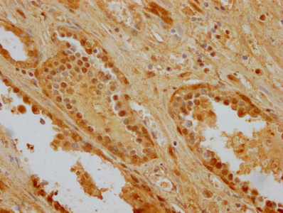
IHC image of CSB-MA010829A0m diluted at 1:256 and staining in paraffin-embedded human prostate cancer performed on a Leica BondTM system. After dewaxing and hydration, antigen retrieval was mediated by high pressure in a citrate buffer (pH 6.0). Section was blocked with 10% normal goat serum 30min at RT. Then primary antibody (1% BSA) was incubated at 4°C overnight, and detected by a Goat anti-mouse IgG polymer labeled by HRP and visualized using 0.05% DAB.
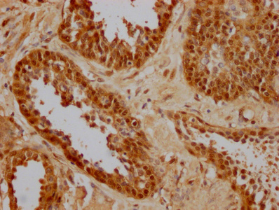
IHC image of CSB-MA010829A0m diluted at 1:256 and staining in paraffin-embedded human prostate cancer performed on a Leica BondTM system. After dewaxing and hydration, antigen retrieval was mediated by high pressure in a citrate buffer (pH 6.0). Section was blocked with 10% normal goat serum 30min at RT. Then primary antibody (1% BSA) was incubated at 4°C overnight, and detected by a Goat anti-mouse IgG polymer labeled by HRP and visualized using 0.05% DAB.
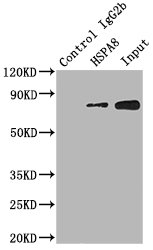
Immunoprecipitating HSPA8 in Hela whole cell lysate
Lane 1: Mouse control IgG2b instead of CSB-MA010829A0m in Hela whole cell lysate
Lane 2: CSB-MA010829A0m (1.5µl) + Hela whole cell lysate (500µg)
Lane 3: Hela whole cell lysate (20µg)
For western blotting, the blot was detected with CSB-MA010829A0m at 1:2000, and a HRP-conjugated Protein G antibody was used as the secondary antibody at 1:2000
.
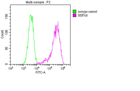
Overlay histogram showing MCF-7 cells stained with CSB-MA010829A0m (red line). The cells were fixed with 70% Ethylalcohol (18h) and then incubated in 10% normal goat serum to block non-specific protein-protein interactions followed by the primary antibody at 1:200 for 1 h at 4°C. The secondary antibody used was FITC goat anti-mouse IgG(H+L) at 1/100 dilution for 30min at 4°C. Isotype control antibody (green line) was mouse IgG2b used under the same conditions. Acquisition of >10,000 events was performed.
The HSPA8 monoclonal antibody is produced from the hybridoma fused by the myeloma cells and splenocytes that were extracted from the immunized mouse. The mouse was immunized with the recombinant human HSPA8 protein (2-646aa). This HSPA8 monoclonal antibody is purified through protein A with a purity of more than 95%. It reacts with samples containing HSPA8 from human, rat, and mouse. And it has been amenable for ELISA, WB, IHC, FC, and IP applications.
HSPA8, also known as Hsc70, mainly acts as a molecular chaperone, which helps to fold newly synthesized proteins, repair misfolded proteins, and target damaged proteins for degradation. It is involved in many cellular processes, including protein folding, assembly and transport, endocytosis, signal transduction, and apoptosis. HSPA8 also plays a critical role in protecting cells from stress-induced damage by stabilizing denatured proteins and preventing protein aggregation.


Email: support@cusabio.com
Distributors Worldwide