Call us
301-363-4651 (Available 9 a.m. to 5 p.m. CST from Monday to Friday)
| Application | Recommended Dilution |
|---|---|
| WB | 1:5000-1:32000 |
| IHC | 1:100-1:500 |
| IF | 1:100-1:500 |
| FC | 1:100-1:300 |
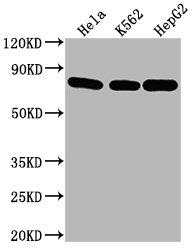
Western Blot
Positive WB detected in: Hela whole cell lysate, K562 whole cell lysate, HepG2 whole cell lysate
All lanes HSPA8 antibody at 1:2000
Secondary
Goat polyclonal to mouse IgG at 1/50000 dilution
Predicted band size: 70~75 KDa
Observed band size: 70~75 KDa
Exposure time: 5min
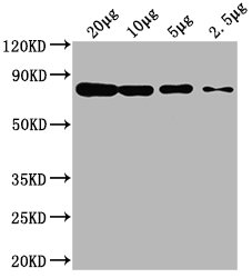
Western Blot
Positive WB detected in: Hela whole cell lysate at 20μg, 10μg, 5μg, 2.5μg
All lanes: HSPA8 antibody at 1:2000
Secondary
Goat polyclonal to mouse IgG at 1/50000 dilution
Predicted band size: 70~75 KDa
Observed band size: 70~75 KDa
Exposure time: 5min
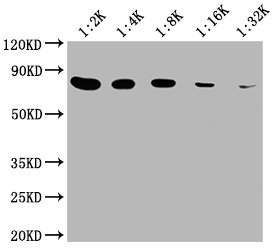
Western Blot
Positive WB detected in: 20μg Hela whole cell lysate
HSPA8 antibody at 1:2000, 1:4000, 1:8000, 1:16000, 1:32000
Secondary
Goat polyclonal to mouse IgG at 1/50000 dilution
Predicted band size: 70~75 KDa
Observed band size: 70~75 KDa
Exposure time: 5min
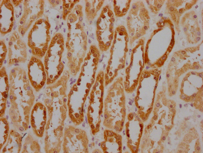
IHC image of CSB-MA010829A1m diluted at 1:220 and staining in paraffin-embedded human kidney tissue performed on a Leica BondTM system. After dewaxing and hydration, antigen retrieval was mediated by high pressure in a citrate buffer (pH 6.0). Section was blocked with 10% normal goat serum 30min at RT. Then primary antibody (1% BSA) was incubated at 4°C overnight. The primary is detected by a biotinylated secondary antibody and visualized using an HRP conjugated SP system.
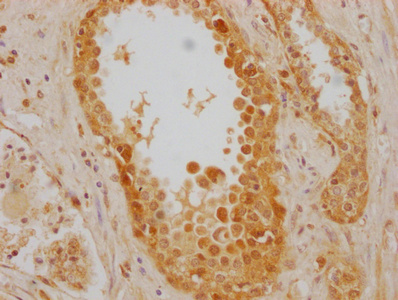
IHC image of CSB-MA010829A1m diluted at 1:220 and staining in paraffin-embedded human prostate cancer performed on a Leica BondTM system. After dewaxing and hydration, antigen retrieval was mediated by high pressure in a citrate buffer (pH 6.0). Section was blocked with 10% normal goat serum 30min at RT. Then primary antibody (1% BSA) was incubated at 4°C overnight. The primary is detected by a biotinylated secondary antibody and visualized using an HRP conjugated SP system.
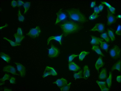
Immunofluorescence staining of Hela cells with CSB-MA010829A1m at 1:100, counter-stained with DAPI. The cells were fixed in 4% formaldehyde and blocked in 10% normal Goat Serum. The cells were then incubated with the antibody overnight at 4°C. Nuclear DNA was labeled in blue with DAPI. The secondary antibody was FITC-conjugated AffiniPure Goat Anti-Mouse IgG (H+L).
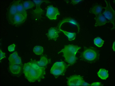
Immunofluorescence staining of MCF-7 cells with CSB-MA010829A1m at 1:100, counter-stained with DAPI. The cells were fixed in 4% formaldehyde and blocked in 10% normal Goat Serum. The cells were then incubated with the antibody overnight at 4°C. Nuclear DNA was labeled in blue with DAPI. The secondary antibody was FITC-conjugated AffiniPure Goat Anti-Mouse IgG (H+L).
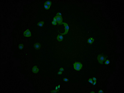
Immunofluorescence staining of PC-3 cells with CSB-MA010829A1m at 1:100, counter-stained with DAPI. The cells were fixed in 4% formaldehyde and blocked in 10% normal Goat Serum. The cells were then incubated with the antibody overnight at 4°C. Nuclear DNA was labeled in blue with DAPI. The secondary antibody was FITC-conjugated AffiniPure Goat Anti-Mouse IgG (H+L).
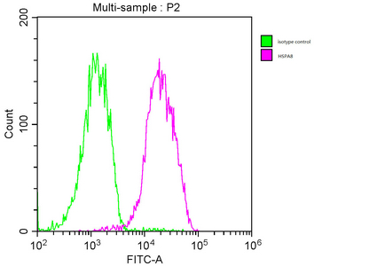
Overlay histogram showing MCF-7 cells stained with CSB-MA010829A1m (red line). The cells were fixed with 70% Ethylalcohol (18h) and then incubated in 10% normal goat serum to block non-specific protein-protein interactions followed by the primary antibody at 1:200 for 1 h at 4°C. The secondary antibody used was FITC goat anti-mouse IgG(H+L) at 1/100 dilution for 30min at 4°C. Isotype control antibody (green line) was mouse IgG1 used under the same conditions. Acquisition of >10,000 events was performed.
This HSPA8 monoclonal antibody was developed by hybridoma technology. The HSPA8 antibody-generating hybridomas are formed by the fusion of the myeloma cells and spleen cells isolated from immunized mice. The mouse is immunized with the recombinant human HSPA8 protein (2-646aa). This HSPA8 monoclonal antibody enables to detect endogenous levels of human HSPA8 protein. It is purified by protein G and quality validated in ELISA, WB, IHC, IF, and FC applications. Its purity is above 95%.
The HSPA8 protein mainly functions as a chaperone in the cell, assisting in protein folding, transport, and degradation by binding to nascent polypeptide chains or misfolded proteins and guiding them through various cellular processes. It plays a crucial role in maintaining cellular homeostasis by preventing the accumulation of misfolded or aggregated proteins that can lead to cellular dysfunction and disease. It is also involved in the regulation of apoptosis, antigen processing, and cell signaling.


Email: support@cusabio.com
Distributors Worldwide