
Call us
301-363-4651 (Available 9 a.m. to 5 p.m. CST from Monday to Friday)

| Application | Recommended Dilution |
|---|---|
| WB | 1:500-1:5000 |
| IHC | 1:50-1:500 |
| IF | 1:50-1:200 |

Western Blot
Positive WB detected in: Jurkat whole cell lysate, THP-1 whole cell lysate, Raji whole cell lysate
All lanes: CD31 antibody at 2.5µg/ml
Secondary
Goat polyclonal to Mouse IgG at 1/50000 dilution
Predicted band size: 83, 81, 80, 82 kDa
Observed band size: 130 kDa
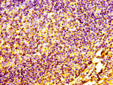
IHC image of CSB-MA017767A0m diluted at 1:100 and staining in paraffin-embedded human tonsil tissue performed on a Leica BondTM system. After dewaxing and hydration, antigen retrieval was mediated by high pressure in a citrate buffer (pH 6.0). Section was blocked with 10% normal goat serum 30min at RT. Then primary antibody (1% BSA) was incubated at 4°C overnight. The primary is detected by a biotinylated secondary antibody and visualized using an HRP conjugated SP system.
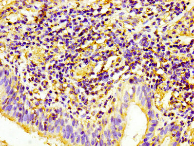
IHC image of CSB-MA017767A0m diluted at 1:100 and staining in paraffin-embedded human lung cancer performed on a Leica BondTM system. After dewaxing and hydration, antigen retrieval was mediated by high pressure in a citrate buffer (pH 6.0). Section was blocked with 10% normal goat serum 30min at RT. Then primary antibody (1% BSA) was incubated at 4°C overnight. The primary is detected by a biotinylated secondary antibody and visualized using an HRP conjugated SP system.
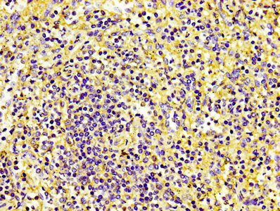
IHC image of CSB-MA017767A0m diluted at 1:100 and staining in paraffin-embedded human spleen tissue performed on a Leica BondTM system. After dewaxing and hydration, antigen retrieval was mediated by high pressure in a citrate buffer (pH 6.0). Section was blocked with 10% normal goat serum 30min at RT. Then primary antibody (1% BSA) was incubated at 4°C overnight. The primary is detected by a biotinylated secondary antibody and visualized using an HRP conjugated SP system.
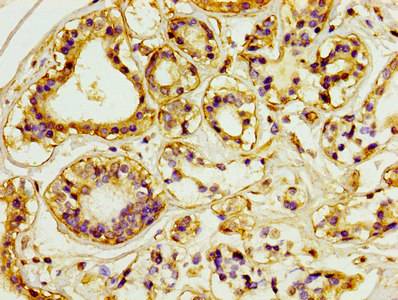
IHC image of CSB-MA017767A0m diluted at 1:100 and staining in paraffin-embedded human breast cancer performed on a Leica BondTM system. After dewaxing and hydration, antigen retrieval was mediated by high pressure in a citrate buffer (pH 6.0). Section was blocked with 10% normal goat serum 30min at RT. Then primary antibody (1% BSA) was incubated at 4°C overnight. The primary is detected by a biotinylated secondary antibody and visualized using an HRP conjugated SP system.
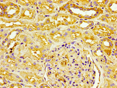
IHC image of CSB-MA017767A0m diluted at 1:100 and staining in paraffin-embedded human kidney tissue performed on a Leica BondTM system. After dewaxing and hydration, antigen retrieval was mediated by high pressure in a citrate buffer (pH 6.0). Section was blocked with 10% normal goat serum 30min at RT. Then primary antibody (1% BSA) was incubated at 4°C overnight. The primary is detected by a biotinylated secondary antibody and visualized using an HRP conjugated SP system.
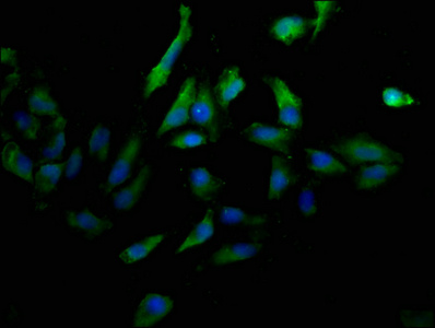
Immunofluorescence staining of Hela cells with CSB-MA017767A0m at 1:100, counter-stained with DAPI. The cells were blocked in 10% normal Goat Serum and then incubated with the primary antibody overnight at 4°C. The secondary antibody was Alexa Fluor 488-congugated AffiniPure Goat Anti-Mouse IgG(H+L).
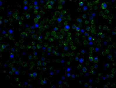
Immunofluorescence staining of THP-1 cells with CSB-MA017767A0m at 1:100, counter-stained with DAPI. The cells were blocked in 10% normal Goat Serum and then incubated with the primary antibody overnight at 4°C. The secondary antibody was Alexa Fluor 488-congugated AffiniPure Goat Anti-Mouse IgG(H+L).
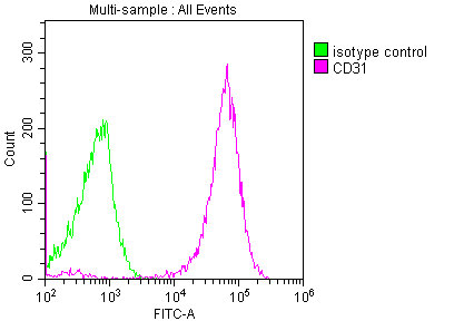
Overlay histogram showing THP-1 cells stained with CSB-MA017767A0m (red line) at 1:250. The cells were incubated in 1x PBS /10% normal goat serum to block non-specific protein-protein interactions followed by primary antibody for 1 h at 4°C. The secondary antibody used was FITC goat anti-mouse IgG(H+L) at 1/200 dilution for 1 h at 4°C. Isotype control antibody (green line) was used under the same conditions. Acquisition of >10,000 events was performed.
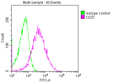
Overlay histogram showing HL-60 cells stained with CSB-MA017767A0m (red line) at 1:250. The cells were incubated in 1x PBS /10% normal goat serum to block non-specific protein-protein interactions followed by primary antibody for 1 h at 4°C. The secondary antibody used was FITC goat anti-mouse IgG(H+L) at 1/200 dilution for 1 h at 4°C. Isotype control antibody (green line) was used under the same conditions. Acquisition of >10,000 events was performed.
The antibody against CD31 was raised in mouse using a portion of the human CD31 extracellular domain (28-315aa) as the immunogen. The antibody exists as a non-conjugated isotype IgG2a, purified by protein A with a purity greater than 95%. This antibody has been validated on ELISA, Western blot (WB), Immunohistochemistry (IHC), Immunofluorescence (IF), and Flow Cytometry (FC). Since PECAM1 or CD31 is related to pro-inflammatory processes and has related roles in angiogenesis, it can be found on carcinomas modulating the immune response. The Platelet endothelial cell adhesion molecule, PECAM1 or CD31, is a protein required for the leukocyte transendothelial migration (TEM). Also, PECAM1 controls the macrophage-mediated phagocytosis of leukocytes. This protein is present in viable leukocytes and platelets and generally concentrated between their borders with epithelial cells. PECAM1 presence makes leukocytes avoid the macrophage phagocytosis with a detachment signaling.
Applications :
Sample type:
Sample dilution:
Review:
By Anonymous


Email: support@cusabio.com
Distributors Worldwide



