
Call us
301-363-4651 (Available 9 a.m. to 5 p.m. CST from Monday to Friday)

| Application | Recommended Dilution |
|---|---|
| WB | 1:5000-1:1600000 |
| IHC | 1:50-1:500 |
| IF | 1:50-1:200 |
| IP | 1µ |
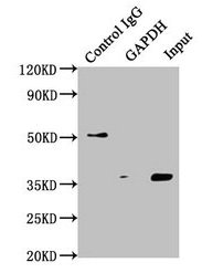
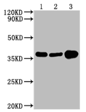
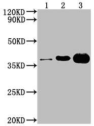
Immunoprecipitating GAPDH in Hela whole cell lysate
Lane 1: Mouse control IgG instead of CSB-MA000071M0m in Hela whole cell lysate.
Lane 2: CSB-MA000071M0m (1μl) + Hela whole cell lysate (500μg)
Lane 3: Hela whole cell lysate (20μg)
For western blotting, the blot was detected with CSB-MA000071M0m at 1:5000, and a HRP-conjugated Protein G antibody was used as the secondary antibody at 1:2000
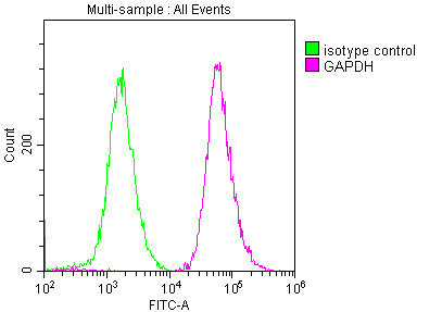
Overlay histogram showing Hela cells stained with CSB-MA000071M0m (red line). The cells were fixed with 70% Ethylalcohol (18h) and then permeabilized with 0.3% Triton X-100 for 2 min. The cells were then incubated in 10% normal goat serum to block non-specific protein-protein interactions followed by the antibody (1:200/1*106cells) for 1 h at 4°C. The secondary antibody used was FITC goat anti-mouse IgG(H+L) at 1/100 dilution for 30min at 4°C. Isotype control antibody (green line) was mouse IgG1 (1:200/1*106cells) used under the same conditions. Acquisition of >10,000 events was performed.
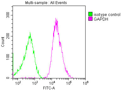
Overlay histogram showing Jurkat cells stained with CSB-MA000071M0m (red line). The cells were fixed with 70% Ethylalcohol (18h) and then permeabilized with 0.3% Triton X-100 for 2 min. The cells were then incubated in 10% normal goat serum to block non-specific protein-protein interactions followed by the antibody (1:200/1*106cells) for 1 h at 4°C. The secondary antibody used was FITC goat anti-mouse IgG(H+L) at 1/100 dilution for 30min at 4°C. Isotype control antibody (green line) was mouse IgG1 (1:200/1*106cells) used under the same conditions. Acquisition of >10,000 events was performed.
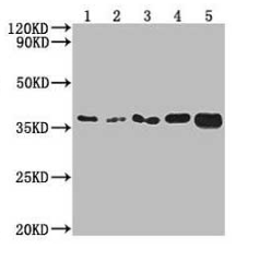
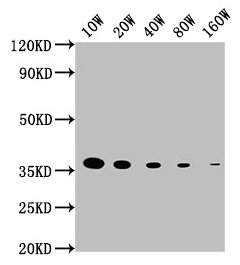
Western Blot
Positive WB detected in: 15μg hela whole cell lysate
GAPDH antibody at 1:100000, 1:200000, 1:400000, 1:800000, 1:1600000
Secondary
Goat polyclonal to mouse IgG at 1/50000 dilution
Predicted band size: 36 KDa
Observed band size: 36 KDa
Exposure time: 5min
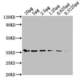
Western Blot
Positive WB detected in: Hela whole cell lysate at 10μg, 5μg, 2.5μg, 1.25μg, 0.625μg, 0.3125μg
All lanes: GAPDH antibody at 1:5000
Secondary
Goat polyclonal to mouse IgG at 1/50000 dilution
Predicted band size: 36 KDa
Observed band size: 36 KDa
Exposure time: 5min
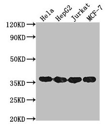
Western Blot
Positive WB detected in: Hela whole cell lysate, HepG2 whole cell lysate, Jurkat whole cell lysate, MCF-7 whole cell lysate
All lanes: GAPDH antibody at 1:2000
Secondary
Goat polyclonal to mouse IgG at 1/50000 dilution
Predicted band size: 36 KDa
Observed band size: 36 KDa
Exposure time: 30s
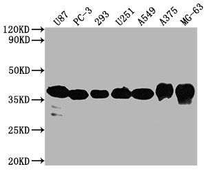
Western Blot
Positive WB detected in: U87 whole cell lysate, PC3 whole cell lysate, 293 whole cell lysate, U251 whole cell lysate, A549 whole cell lysate, A375 whole cell lysate, MG-63 whole cell lysate
All lanes GAPDH antibody at 1:5000
Secondary
Goat polyclonal to mouse IgG at 1/50000 dilution
Predicted band size: 36 KDa
Observed band size: 36 KDa
Exposure time: 30s
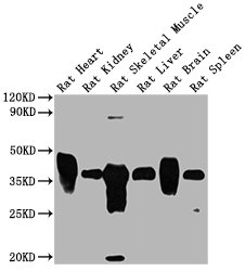
Western Blot
Positive WB detected in: Rat heart tissue, Rat kidney tissue, Rat skeletal muscle tissue, Rat liver tissue, Rat brain tissue tissue, Rat spleen tissue
All lanes GAPDH antibody at 1:1000
Secondary
Goat polyclonal to mouse IgG at 1/50000 dilution
Predicted band size: 36 KDa
Observed band size: 36 KDa
Exposure time: 1min
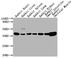
Western Blot
Positive WB detected in: Rabbit heart tissue, Rabbit liver tissue,Rabbit spleen tissue, Rabbit lung tissue, Rabbit kidney tissue, Rabbit small intestine tissue, Rabbit skeletal muscle tissue
All lanes GAPDH antibody at 1:5000
Secondary
Goat polyclonal to mouse IgG at 1/50000 dilution
Predicted band size: 36 KDa
Observed band size: 36 KDa
Exposure time: 5min
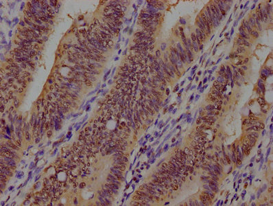
IHC image of CSB-MA000071M0m diluted at 1:100 and staining in paraffin-embedded human colon cancer performed on a Leica BondTM system. After dewaxing and hydration, antigen retrieval was mediated by high pressure in a citrate buffer (pH 6.0). Section was blocked with 10% normal goat serum 30min at RT. Then primary antibody (1% BSA) was incubated at 4°C overnight. The primary is detected by a biotinylated secondary antibody and visualized using an HRP conjugated SP system.
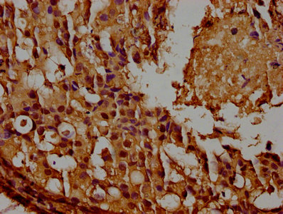
IHC image of CSB-MA000071M0m diluted at 1:100 and staining in paraffin-embedded human breast cancer performed on a Leica BondTM system. After dewaxing and hydration, antigen retrieval was mediated by high pressure in a citrate buffer (pH 6.0). Section was blocked with 10% normal goat serum 30min at RT. Then primary antibody (1% BSA) was incubated at 4°C overnight. The primary is detected by a biotinylated secondary antibody and visualized using an HRP conjugated SP system.
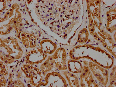
IHC image of CSB-MA000071M0m diluted at 1:100 and staining in paraffin-embedded human kidney tissue performed on a Leica BondTM system. After dewaxing and hydration, antigen retrieval was mediated by high pressure in a citrate buffer (pH 6.0). Section was blocked with 10% normal goat serum 30min at RT. Then primary antibody (1% BSA) was incubated at 4°C overnight. The primary is detected by a biotinylated secondary antibody and visualized using an HRP conjugated SP system.
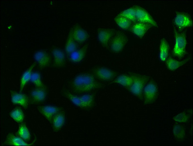
Immunofluorescence staining of Hela cells with CSB-MA000071M0m at 1:220, counter-stained with DAPI. The cells were fixed in 4% formaldehyde, permeabilized using 0.2% Triton X-100 and blocked in 10% normal Goat Serum. The cells were then incubated with the antibody overnight at 4°C. Nuclear DNA was labeled in blue with DAPI. The secondary antibody was FITC-conjugated AffiniPure Goat Anti-Mouse IgG(H+L).
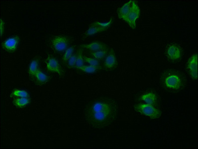
Immunofluorescence staining of HepG2 cells with CSB-MA000071M0m at 1:220, counter-stained with DAPI. The cells were fixed in 4% formaldehyde, permeabilized using 0.2% Triton X-100 and blocked in 10% normal Goat Serum. The cells were then incubated with the antibody overnight at 4°C. Nuclear DNA was labeled in blue with DAPI. The secondary antibody was FITC-conjugated AffiniPure Goat Anti-Mouse IgG(H+L).
The GAPDH Monoclonal Antibody is a specific antibody that targets GAPDH. The GAPDH antibody is an internal reference antibody that functions as a loading control to ensure equal protein loading and accurate quantification of protein expression levels in different samples. This antibody can detect GAPDH in human, mouse, and rabbit species.
The immunogen used to generate this GAPDH antibody is the 2-335 amino acid region of recombinant Human GAPDH protein. The GAPDH Monoclonal Antibody is raised in mouse and belongs to the IgG1 isotype. It is purified using Protein G and reaches a purity level of greater than 95%.
The GAPDH Monoclonal Antibody is available in liquid form and has been tested in various applications, including ELISA, WB, IHC, IP, and IF. These applications make the antibody a versatile tool for the detection and analysis of GAPDH in different contexts.
Moreover, the GAPDH Monoclonal Antibody has been cited in a paper by H Miao, et al. in 2022, which highlights its utility in scientific research. The use of this validated antibody in research increases the reliability of the results and ensures reproducibility.
Applications :
Sample type:
Sample dilution:
Review:
By Anonymous
Applications :
Sample type:
Sample dilution:
Review:
By Anonymous
Applications :
Sample type:
Sample dilution:
Review:
By Anonymous
Applications :
Sample type:
Sample dilution:
Review:
By Anonymous
Applications :
Sample type:
Sample dilution:
Review:
By Anonymous
Applications :
Sample type:
Sample dilution:
Review:
By Anonymous

Applications :
Sample type:
Sample dilution:
Review:
By Anonymous
Applications :
Review:
By Anonymous
Applications :
Sample dilution:
Review:
By Anonymous
Applications :
Sample dilution:
Review:
By Anonymous
Applications :
Sample dilution:
Review:
By Anonymous


Email: support@cusabio.com
Distributors Worldwide



