Recombinant Human HLA class II histocompatibility antigen, DR beta 5 chain (HLA-DRB5), partial

In Stock
Code: CSB-EP657235HU
Size:
20ug
20ug100ug1mg(1mg*1 or 500ug*2)
US$388
Quantity:
Express system: E.coli
Species: Homo sapiens (Human)
Tag Info: N-terminal 6xHis-tagged
For inquiries on large quantities or another requirements
Send an Inquiry
Start an on-line Chat
Online ordering is currently available for U.S. customers only. For orders outside the U.S., please kindly submit an inquiry or start a chat with us.

Product Details

Purity
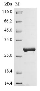
Greater than 90% as determined by SDS-PAGE.
Target Names
HLA-DRB5
Uniprot NO.
Research Area
Immunology
Species
Homo sapiens (Human)
Source
E.coli
Expression Region
30-227aa
Target Protein Sequence
GDTRPRFLQQDKYECHFFNGTERVRFLHRDIYNQEEDLRFDSDVGEYRAVTELGRPDAEYWNSQKDFLEDRRAAVDTYCRHNYGVGESFTVQRRVEPKVTVYPARTQTLQHHNLLVCSVNGFYPGSIEVRWFRNSQEEKAGVVSTGLIQNGDWTFQTLVMLETVPRSGEVYTCQVEHPSVTSPLTVEWRAQSESAQSK
Note: The complete sequence may include tag sequence, target protein sequence, linker sequence and extra sequence that is translated with the protein sequence for the purpose(s) of secretion, stability, solubility, etc.
If the exact amino acid sequence of this recombinant protein is critical to your application, please explicitly request the full and complete sequence of this protein before ordering.
Mol. Weight
27.1 kDa
Protein Length
Partial
Tag Info
N-terminal 6xHis-tagged
The tag type will be determined during production process. If you have specified tag type, please tell us and we will develop the specified tag preferentially.
Form
Liquid or Lyophilized powder
Note: We will preferentially ship the format that we have in stock, however, if you have any special requirement for the format, please remark your requirement when placing the order, we will prepare according to your demand.
Buffer
If the delivery form is liquid, the default storage buffer is Tris/PBS-based buffer, 5%-50% glycerol. If the delivery form is lyophilized powder, the buffer before lyophilization is Tris/PBS-based buffer, 6% Trehalose.
Reconstitution
We recommend that this vial be briefly centrifuged prior to opening to bring the contents to the bottom. Please reconstitute protein in deionized sterile water to a concentration of 0.1-1.0 mg/mL.We recommend to add 5-50% of glycerol (final concentration) and aliquot for long-term storage at -20°C/-80°C. Our default final concentration of glycerol is 50%. Customers could use it as reference.
Storage
The shelf life is related to many factors, storage state, buffer ingredients, storage temperature and the stability of the protein itself. Generally, the shelf life of liquid form is 6 months at -20°C/-80°C. The shelf life of lyophilized form is 12 months at -20°C/-80°C.
Shelf Life
The shelf life is related to many factors, storage state, buffer ingredients, storage temperature and the stability of the protein itself.
Generally, the shelf life of liquid form is 6 months at -20°C/-80°C. The shelf life of lyophilized form is 12 months at -20°C/-80°C.
Lead Time
3-7 business days
Note: All of our proteins are default shipped with normal blue ice packs, if you request to ship with dry ice, please communicate with us in advance and extra fees will be charged.
Notes
Repeated freezing and thawing is not recommended. Store working aliquots at 4°C for up to one week.
Troubleshooting and FAQs
Datasheet & COA
Please contact us to get it.
Images
  • (Tris-Glycine gel) Discontinuous SDS-PAGE (reduced) with 5% enrichment gel and 15% separation gel.

Customer Reviews and Q&A

 Customer Reviews

Target Background

Function(From Uniprot)
Binds peptides derived from antigens that access the endocytic route of antigen presenting cells (APC) and presents them on the cell surface for recognition by the CD4 T-cells. The peptide binding cleft accommodates peptides of 10-30 residues. The peptides presented by MHC class II molecules are generated mostly by degradation of proteins that access the endocytic route, where they are processed by lysosomal proteases and other hydrolases. Exogenous antigens that have been endocytosed by the APC are thus readily available for presentation via MHC II molecules, and for this reason this antigen presentation pathway is usually referred to as exogenous. As membrane proteins on their way to degradation in lysosomes as part of their normal turn-over are also contained in the endosomal/lysosomal compartments, exogenous antigens must compete with those derived from endogenous components. Autophagy is also a source of endogenous peptides, autophagosomes constitutively fuse with MHC class II loading compartments. In addition to APCs, other cells of the gastrointestinal tract, such as epithelial cells, express MHC class II molecules and CD74 and act as APCs, which is an unusual trait of the GI tract. To produce a MHC class II molecule that presents an antigen, three MHC class II molecules (heterodimers of an alpha and a beta chain) associate with a CD74 trimer in the ER to form a heterononamer. Soon after the entry of this complex into the endosomal/lysosomal system where antigen processing occurs, CD74 undergoes a sequential degradation by various proteases, including CTSS and CTSL, leaving a small fragment termed CLIP (class-II-associated invariant chain peptide). The removal of CLIP is facilitated by HLA-DM via direct binding to the alpha-beta-CLIP complex so that CLIP is released. HLA-DM stabilizes MHC class II molecules until primary high affinity antigenic peptides are bound. The MHC II molecule bound to a peptide is then transported to the cell membrane surface. In B-cells, the interaction between HLA-DM and MHC class II molecules is regulated by HLA-DO. Primary dendritic cells (DCs) also to express HLA-DO. Lysosomal microenvironment has been implicated in the regulation of antigen loading into MHC II molecules, increased acidification produces increased proteolysis and efficient peptide loading.
Gene References into Functions
  1. Variation at HLA-DRB5 being associated both with Parkinson's disease and Alzheimer's disease, ariation at HLA-DRB5 being associated both with Parkinson's disease and Alzheimer's disease. PMID:27713094
  2. This study identified genetic overlap between Alzheimer disease and immune-mediated diseases, implicating the HLA locus and IPMK in the pathobiology of Alzheimer disease. PMID:27088644
  3. HLA-DRB5 affects type 1 diabetes risk and islet autoantibodies. PMID:26740600
  4. Brain DNA methylation in HLA-DRB5 was associated with pathological Alzheimer disease. PMID:25365775
  5. findings indicated that copy number variants of HLA-DRB5 was associated with the risk of systemic lupus erythematosus, and copy number deletion appeared to be protective for SLE. PMID:24366815
  6. DRB5*01:01 is associated with Thai systemic lupus erythematosus; the association is stronger than that of DRB1*15:01; genetic contribution of DRB5*01:01 is due partially to linkage disequilibrium between DRB1*16:02 and DRB5*01:01 in northern Thai population PMID:22862923
  7. HLA-DRB5 was highly expressed in peripheral blood mononuclear cells from patients with systemic sclerosis-related interstitial lung disease. The HLA-DRB5*01:05 allele is a risk factor for interstitial lung disease in patients with systemic sclerosis. PMID:22723597
  8. Transgenic mice expressing HLA-DRB5*01:01 genes are crossed with histocompatibility class (MHC) II knockout mice to replace mouse MHC class II genes with human MHC class II genes in a humanized model of autoimmunity. PMID:22888134
  9. Eighteen out of 19 individuals showed HLA-DRB5, and none of the HLA-DRB5*null individuals, carried the HLADRB1* 15 allele. The findings support a contribution for HLA-DRB5 in keloid pathogenesis. PMID:22033527
  10. HLA-DRB5 was found to have bimodal expression in human skeletal muscle tissue. PMID:21299892
  11. In our study, we found that the HLA-DR9 allele and HPV16E6 infection had a function of synergy in the process of malignant transformation of esophageal epithelial cells, and jointly promoting the occurrence and development of esophageal cancer. PMID:20193235
  12. the identification of a new HLA-DRB5 allele found in two members of a British Caucasoid family. PMID:14617041
  13. hMBP82-100-specific type B T cells escaped tolerance in HLA-DRB5*0101 Tg mice PMID:18713991
  14. the gene frequency of HLADR5 was significantly decreased in the total group of patients with autoimmune liver disease PMID:19811438

Show More

Hide All

Subcellular Location
Cell membrane; Single-pass type I membrane protein. Endoplasmic reticulum membrane; Single-pass type I membrane protein. Golgi apparatus, trans-Golgi network membrane; Single-pass type I membrane protein. Endosome membrane; Single-pass type I membrane protein. Lysosome membrane; Single-pass type I membrane protein. Late endosome membrane; Single-pass type I membrane protein. Note=The MHC class II complex transits through a number of intracellular compartments in the endocytic pathway until it reaches the cell membrane for antigen presentation.
Protein Families
MHC class II family
Database Links

HGNC: 4953

UNIGENE: Hs.485130

KEGG: hsa:3127

STRING: 9606.ENSP00000364114

OMIM: 604776

icon of phone
Call us
301-363-4651 (Available 9 a.m. to 5 p.m. CST from Monday to Friday)
icon of address
Address
No. 269, Shendun 5th Road, Donghu Hi-Tech Development Area, Hubei Province, 430206, P.R.China
icon of social media
Join us with

Subscribe newsletter

Leave a message

* To protect against spam, please pass the CAPTCHA test below.
CAPTCHA verification
© 2007-2025 CUSABIO TECHNOLOGY LLC All rights reserved. 鄂ICP备15011166号-1
Select 0 Products