
Call us
301-363-4651 (Available 9 a.m. to 5 p.m. CST from Monday to Friday)

| Application | Recommended Dilution |
|---|---|
| IHC | 1:20-1:200 |
| IF | 1:50-1:200 |
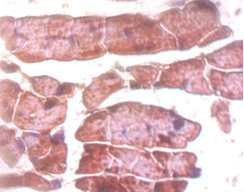
Immunohistochemistry of paraffin-embedded mouse heart tissue using CSB-PA15079A0Rb at dilution of 1:50
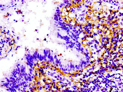
IHC image of CSB-PA15079A0Rb diluted at 1:100 and staining in paraffin-embedded human lung cancer performed on a Leica BondTM system. After dewaxing and hydration, antigen retrieval was mediated by high pressure in a citrate buffer (pH 6.0). Section was blocked with 10% normal goat serum 30min at RT. Then primary antibody (1% BSA) was incubated at 4°C overnight. The primary is detected by a biotinylated secondary antibody and visualized using an HRP conjugated SP system.
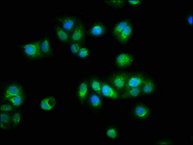
Immunofluorescence staining of HepG2 cells with CSB-PA15079A0Rb at 1:100, counter-stained with DAPI. The cells were fixed in 4% formaldehyde, permeabilized using 0.2% Triton X-100 and blocked in 10% normal Goat Serum. The cells were then incubated with the antibody overnight at 4°C. The secondary antibody was Alexa Fluor 488-congugated AffiniPure Goat Anti-Rabbit IgG(H+L).
The polyclonal antibody is raised in the rabbit against amino acids 5398-5604 of Titin protein (TTN). It is protein G purified and reaches over 95% in purity. This TTN antibody exists as an unconjugated IgG isoform. It only reacts with human TNN protein. And it has been validated for use in three applications, including ELISA, IHC, and IF. Its target protein TTN is a giant filament protein that plays an important role in skeletal and cardiac muscles. And it also participates in sarcomere formation, mechanosensing, and signal transduction.


Email: support@cusabio.com
Distributors Worldwide



