
Call us
301-363-4651 (Available 9 a.m. to 5 p.m. CST from Monday to Friday)

| Application | Recommended Dilution |
|---|---|
| WB | 1:500-1:5000 |
| IF | 1:200-1:500 |
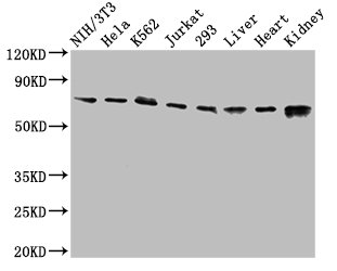
Western Blot
Positive WB detected in: NIH/3T3 whole cell lysate, Hela whole cell lysate, K562 whole cell lysate, Jurkat whole cell lysate, 293 whole cell lysate, Rat liver tissue, Mouse heart tissue, Mouse kidney tissue
All lanes: RIPK1 antibody at 3.2µg/ml
Secondary
Goat polyclonal to rabbit IgG at 1/50000 dilution
Predicted band size: 76, 71 kDa
Observed band size: 76 kDa
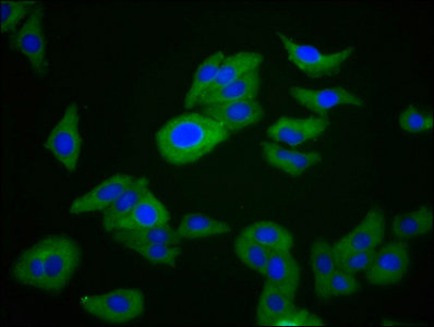
Immunofluorescence staining of HepG2 cells with CSB-PA618785LA01HU at 1:266, counter-stained with DAPI. The cells were fixed in 4% formaldehyde, permeabilized using 0.2% Triton X-100 and blocked in 10% normal Goat Serum. The cells were then incubated with the antibody overnight at 4°C. The secondary antibody was Alexa Fluor 488-congugated AffiniPure Goat Anti-Rabbit IgG(H+L).
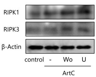
Applications :
Review:
By Anonymous


Email: support@cusabio.com
Distributors Worldwide



