
Call us
301-363-4651 (Available 9 a.m. to 5 p.m. CST from Monday to Friday)

| Application | Recommended Dilution |
|---|---|
| WB | 1:1000-1:5000 |
| IHC | 1:500-1:1000 |
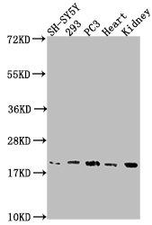
Western Blot
Positive WB detected in: SH-SY5Y whole cell lysate, 293 whole cell lysate, PC3 whole cell lysate, Mouse heart tissue, Mouse kidney tissue
All lanes: PYCARD antibody at 1:2000
Secondary
Goat polyclonal to rabbit IgG at 1/50000 dilution
Predicted band size: 22, 20, 16 kDa
Observed band size: 20 kDa
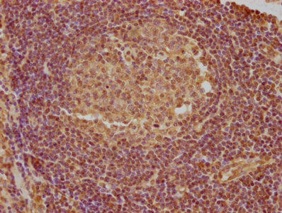
IHC image of CSB-PA890936LA01HU diluted at 1:740 and staining in paraffin-embedded human lymph node tissue performed on a Leica BondTM system. After dewaxing and hydration, antigen retrieval was mediated by high pressure in a citrate buffer (pH 6.0). Section was blocked with 10% normal goat serum 30min at RT. Then primary antibody (1% BSA) was incubated at 4°C overnight. The primary is detected by a biotinylated secondary antibody and visualized using an HRP conjugated SP system.
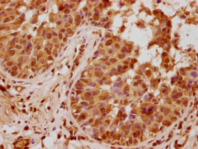
IHC image of CSB-PA890936LA01HU diluted at 1:740 and staining in paraffin-embedded human breast cancer performed on a Leica BondTM system. After dewaxing and hydration, antigen retrieval was mediated by high pressure in a citrate buffer (pH 6.0). Section was blocked with 10% normal goat serum 30min at RT. Then primary antibody (1% BSA) was incubated at 4°C overnight. The primary is detected by a biotinylated secondary antibody and visualized using an HRP conjugated SP system.


Email: support@cusabio.com
Distributors Worldwide



