OPN1SW Antibody

Code: CSB-PA016354LA01HU
Size:
20μg
20μg50μg100μg
US$83
Quantity:
Species Reactivity: Human
Raised in: Rabbit
Application: ELISA, WB, IF
For inquiries on large quantities or another requirements
Send an Inquiry
Start an on-line Chat
Online ordering is currently available for U.S. customers only. For orders outside the U.S., please kindly submit an inquiry or start a chat with us.

Product Details

Uniprot NO.
Target Names
OPN1SW
Alternative Names
OPN1SW; BCP; Short-wave-sensitive opsin 1; Blue cone photoreceptor pigment; Blue-sensitive opsin; BOP
Raised in
Rabbit
Species Reactivity
Human
Immunogen
Recombinant Human Short-wave-sensitive opsin 1 protein (307-348AA)
Immunogen Species
Homo sapiens (Human)
Conjugate
Non-conjugated
Isotype
IgG
Purification Method
>95%, Protein G purified
Concentration
It differs from different batches. Please contact us to confirm it.
Buffer
Preservative: 0.03% Proclin 300
Constituents: 50% Glycerol, 0.01M PBS, pH 7.4
Tested Applications
ELISA, WB, IF
Recommended Dilution
ApplicationRecommended Dilution
WB1:500-1:5000
IF1:50-1:200
Storage
Upon receipt, store at -20°C or -80°C. Avoid repeated freeze.
Lead Time
Basically, we can dispatch the products out in 1-3 working days after receiving your orders. Delivery time maybe differs from different purchasing way or location, please kindly consult your local distributors for specific delivery time.
Note: All of our proteins are default shipped with normal blue ice packs, if you request to ship with dry ice, please communicate with us in advance and extra fees will be charged.
Usage
For Research Use Only. Not for use in diagnostic or therapeutic procedures.
Datasheet & COA
Images
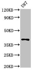
  • Western Blot
    Positive WB detected in: U87 whole cell lysate
    All lanes: OPN1SW antibody at 3μg/ml
    Secondary
    Goat polyclonal to rabbit IgG at 1/50000 dilution
    Predicted band size: 40 kDa
    Observed band size: 40 kDa

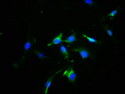
  • Immunofluorescence staining of U251 cells with CSB-PA016354LA01HU at 1:100, counter-stained with DAPI. The cells were fixed in 4% formaldehyde, permeabilized using 0.2% Triton X-100 and blocked in 10% normal Goat Serum. The cells were then incubated with the antibody overnight at 4°C. The secondary antibody was Alexa Fluor 488-congugated AffiniPure Goat Anti-Rabbit IgG(H+L).

Customer Reviews and Q&A

 Customer Reviews
Average Rating:
5.0 - 1 reviews

Submit a Review here

Applications : immunohistochemistry

Sample type: cells

Review: OPN1SW antibodies, which are the rod OS, green/red cones OS, and blue cone markers, respectively. In the MO-injected larvae, the expressions of Rho, ZPR1, and OPN1SW were dramatically suppressed.

By Anonymous

Target Background

Function(From Uniprot)
Visual pigments are the light-absorbing molecules that mediate vision. They consist of an apoprotein, opsin, covalently linked to cis-retinal. Required for the maintenance of cone outer segment organization in the ventral retina, but not essential for the maintenance of functioning cone photoreceptors. Involved in ensuring correct abundance and localization of retinal membrane proteins. May increase spectral sensitivity in dim light.
Gene References into Functions
  1. LVAVA haplotype of the OPN1LW gene and MVAVA haplotype of the OPN1MW gene cause apparently nonsyndromic high myopia in young patients but lead to progressive cone-rod dystrophy with deuteranopia and protanopia in middle-aged patients corresponding to a previously unknown disease course. PMID:28358949
  2. Data suggest that insights into dimerization interface of red cone opsin should aid investigations of the structure and function of GPCR cell signaling. PMID:28045251
  3. A novel homozygous PDE6C mutation was identified as the cause of ACHM. In addition, we identified an OPN1SW mutation in the sibling with complete achromatopsia. PMID:25605338
  4. Individuals with the T190I S-opsin mutation behaved as mild tritans at 12.3-92.3Td, but as tritanopes at 1.2-9.2Td, for both light-adapted and dark-adapted conditions. The results are consistent with the mutant opsin causing abnormal S-cone function. PMID:23022137
  5. A novel mutation(prolin/leucine) in the short-wavelength-sensitive cone pigment gene associated with a tritan color vision defect. PMID:16961973
  6. Results show that, although light absorption behaves differently in blue, green and red opsins, their low-frequency vibrational motions are similar. PMID:19189139
  7. 11-cis-retinol had no significant effect on the activity of human blue cone opsin PMID:19386593
  8. Immunoreactivity to anti-OPN1SW antibodies was seen in the upper layer of human epidermis & reconstructed skin. The opsin mRNA was seen in total RNA from human skin. Neither immunoreactivity nor mRNA expression was seen in cultured human keratinocytes. PMID:19493002

Show More

Hide All

Subcellular Location
Cell membrane; Multi-pass membrane protein. Photoreceptor inner segment. Cell projection, cilium, photoreceptor outer segment. Cytoplasm, perinuclear region.
Protein Families
G-protein coupled receptor 1 family, Opsin subfamily
Tissue Specificity
The three color pigments are found in the cone photoreceptor cells. Expressed throughout the epidermis and dermis, primarily in the stratum granulosum in the facial and abdominal skin (at protein level). Expressed in dermal fibroblasts (at protein level).
Database Links

HGNC: 1012

UNIGENE: Hs.656404

KEGG: hsa:611

STRING: 9606.ENSP00000249389

OMIM: 190900

icon of phone
Call us
301-363-4651 (Available 9 a.m. to 5 p.m. CST from Monday to Friday)

icon of address
Address
No. 269, Shendun 5th Road, Donghu Hi-Tech Development Area, Hubei Province, 430206, P.R.China
icon of social media
Join us with

Subscribe newsletter

Leave a message

* To protect against spam, please pass the CAPTCHA test below.
CAPTCHA verification
© 2007-2025 CUSABIO TECHNOLOGY LLC All rights reserved. 鄂ICP备15011166号-1
Select 0 Products