Call us
301-363-4651 (Available 9 a.m. to 5 p.m. CST from Monday to Friday)
| Application | Recommended Dilution |
|---|---|
| WB | 1:500-1:5000 |
| IF | 1:50-1:200 |
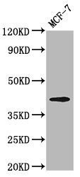
Western Blot
Positive WB detected in: MCF-7 whole cell lysate
All lanes: OPN1MW antibody at 3.2µg/ml
Secondary
Goat polyclonal to rabbit IgG at 1/50000 dilution
Predicted band size: 41 kDa
Observed band size: 41 kDa
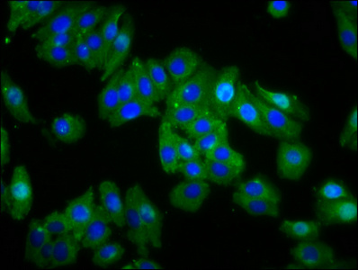
Immunofluorescence staining of HepG2 cells with CSB-PA016352LA01HU at 1:66, counter-stained with DAPI. The cells were fixed in 4% formaldehyde, permeabilized using 0.2% Triton X-100 and blocked in 10% normal Goat Serum. The cells were then incubated with the antibody overnight at 4°C. The secondary antibody was Alexa Fluor 488-congugated AffiniPure Goat Anti-Rabbit IgG(H+L).
Polyclonal rabbit anti-OPN1MW antibody raised against a recombinant protein containing the 1-52 amino acids of the human OPN1MW is recommended to detect the human OPN1MW protein. It was subjected to protein G purification and reached up to 95% in purity. Three applications including ELISA, WB, and IF have been used in assays to test for the specificity of this OPN1MW antibody.
OPN1MW is primarily expressed in the cone cells of the retina, where it is responsible for color vision and visual acuity in bright light conditions. Mutations in the OPN1MW gene have been associated with various visual disorders, including color blindness and macular degeneration.


Email: support@cusabio.com
Distributors Worldwide