MACF1 Antibody

Code: CSB-PA890946LA01HU
Size:
20μl
20μl50μl100μl
US$83
Quantity:
Species Reactivity: Human
Raised in: Rabbit
Application: ELISA, IHC
For inquiries on large quantities or another requirements
Send an Inquiry
Start an on-line Chat
Online ordering is currently available for U.S. customers only. For orders outside the U.S., please kindly submit an inquiry or start a chat with us.

Product Details

Uniprot NO.
Target Names
MACF1
Alternative Names
MACF1 antibody; ABP620 antibody; ACF7 antibody; KIAA0465 antibody; KIAA1251 antibody; Microtubule-actin cross-linking factor 1 antibody; isoforms 1/2/3/5 antibody; 620 kDa actin-binding protein antibody; ABP620 antibody; Actin cross-linking family protein 7 antibody; Macrophin-1 antibody; Trabeculin-alpha antibody
Raised in
Rabbit
Species Reactivity
Human
Immunogen
Recombinant Human Microtubule-actin cross-linking factor 1, isoforms 1/2/3/5 protein (1936-2150AA)
Immunogen Species
Homo sapiens (Human)
Conjugate
Non-conjugated
Isotype
IgG
Purification Method
>95%, Protein G purified
Concentration
It differs from different batches. Please contact us to confirm it.
Buffer
Preservative: 0.03% Proclin 300
Constituents: 50% Glycerol, 0.01M PBS, pH 7.4
Tested Applications
ELISA, IHC
Recommended Dilution
ApplicationRecommended Dilution
IHC1:200-1:500
Storage
Upon receipt, store at -20°C or -80°C. Avoid repeated freeze.
Lead Time
Basically, we can dispatch the products out in 1-4 working days after receiving your orders. Delivery time maybe differs from different purchasing way or location, please kindly consult your local distributors for specific delivery time.
Note: All of our proteins are default shipped with normal blue ice packs, if you request to ship with dry ice, please communicate with us in advance and extra fees will be charged.
Usage
For Research Use Only. Not for use in diagnostic or therapeutic procedures.
Datasheet & COA
Images
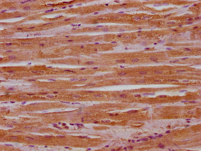
  • IHC image of CSB-PA890946LA01HU diluted at 1:400 and staining in paraffin-embedded human heart tissue performed on a Leica BondTM system. After dewaxing and hydration, antigen retrieval was mediated by high pressure in a citrate buffer (pH 6.0). Section was blocked with 10% normal goat serum 30min at RT. Then primary antibody (1% BSA) was incubated at 4°C overnight. The primary is detected by a biotinylated secondary antibody and visualized using an HRP conjugated SP system.

Customer Reviews and Q&A

 Customer Reviews

Target Background

Function(From Uniprot)
F-actin-binding protein which plays a role in cross-linking actin to other cytoskeletal proteins and also binds to microtubules. Plays an important role in ERBB2-dependent stabilization of microtubules at the cell cortex. Acts as a positive regulator of Wnt receptor signaling pathway and is involved in the translocation of AXIN1 and its associated complex (composed of APC, CTNNB1 and GSK3B) from the cytoplasm to the cell membrane. Has actin-regulated ATPase activity and is essential for controlling focal adhesions (FAs) assembly and dynamics. Interaction with CAMSAP3 at the minus ends of non-centrosomal microtubules tethers microtubules minus-ends to actin filaments, regulating focal adhesion size and cell migration. May play role in delivery of transport vesicles containing GPI-linked proteins from the trans-Golgi network through its interaction with GOLGA4. Plays a key role in wound healing and epidermal cell migration. Required for efficient upward migration of bulge cells in response to wounding and this function is primarily rooted in its ability to coordinate microtubule dynamics and polarize hair follicle stem cells. As a regulator of actin and microtubule arrangement and stabilization, it plays an essential role in neurite outgrowth, branching and spine formation during brain development.
Gene References into Functions
  1. Loss of ACF7 leads to aberrant microtubule organization, tight junction stabilization and impaired wound closure in vitro. ACF7 level is also correlated with development and progression of ulcerative colitis (UC) in patients. PMID:28541346
  2. Study summarized the physiological role of MACF1 as well as its pathological one in various cancers. MACF1 comprises different isoforms, and is broadly expressed in brain, spinal cord, lung, kidney, heart, bone and skeletal muscles tissues. It plays a crucial role in cell proliferation, migration and cell signaling, and is also closely associated with many cancer. PMID:28782898
  3. in mammalian intestinal epithelial cells, the spectraplakin ACF7 (also known as MACF1) specifically binds to CAMSAP3 and is required for the apical localization of CAMSAP3-decorated microtubule minus ends. PMID:27802168
  4. MACF1b may contribute to the genetic etiology and mechanistic causation of Parkinson's disease. PMID:27021023
  5. ACF7, a member of the spectraplakin family of cytoskeletal crosslinking proteins, interacts with Nezha (also called CAMSAP3) at the minus ends of noncentrosomal microtubules and anchors them to actin filaments. PMID:27693509
  6. the present study represents the first investigation on the functional role of MACF1 in tumor cell biology, as well as demonstrates its potential as a unique biomarker that can be targeted synergistically with TMZ as part of a combinatorial therapeutic approach for the treatment of genetically multifarious glioblastomas PMID:27959385
  7. Duplication in the microtubule-actin cross-linking factor 1 gene causes neuromuscular diseases. PMID:24899269
  8. uncovered a role for ELMO in the recruitment of ACF7 to the membrane to promote microtubule capture and stability PMID:23184944
  9. ACF7 targeting to the plasma membrane is both required and sufficient for microtubule capture downstream of ErbB2. PMID:20937854
  10. p230, through its interaction with MACF1, provides the molecular link for transport of GPI-anchored proteins along the microtubule and actin cytoskeleton from the TGN to the cell periphery. PMID:15265687
  11. In two lung cell lines, MACF1b was chiefly localized to the Golgi complex. The domain of MACF1b that targets it to the Golgi was found at the N-terminal part of the region that contains the plakin repeats. PMID:16076900

Show More

Hide All

Subcellular Location
[Isoform 2]: Cytoplasm, cytoskeleton. Cytoplasm. Golgi apparatus. Cell membrane. Cell projection, ruffle membrane.; [Isoform 1]: Cytoplasm. Golgi apparatus.
Protein Families
Plakin or cytolinker family
Tissue Specificity
Isoform 2: Ubiquitously expressed. Isoform 1: Expressed in cell lines NCI-H460, A-549 and HaCaT. Isoform 4: Expressed in heart, lung, pituitary and placenta, not found in brain, kidney, liver, pancreas or skeletal muscle.
Database Links

HGNC: 13664

UNIGENE: Hs.472475

KEGG: hsa:23499

STRING: 9606.ENSP00000354573

OMIM: 608271

icon of phone
Call us
301-363-4651 (Available 9 a.m. to 5 p.m. CST from Monday to Friday)

icon of address
Address
No. 269, Shendun 5th Road, Donghu Hi-Tech Development Area, Hubei Province, 430206, P.R.China
icon of social media
Join us with

Subscribe newsletter

Leave a message

* To protect against spam, please pass the CAPTCHA test below.
CAPTCHA verification
© 2007-2025 CUSABIO TECHNOLOGY LLC All rights reserved. 鄂ICP备15011166号-1
Select 0 Products