Call us
301-363-4651 (Available 9 a.m. to 5 p.m. CST from Monday to Friday)
| Application | Recommended Dilution |
|---|---|
| IHC | 1:20-1:200 |
| IF | 1:50-1:200 |
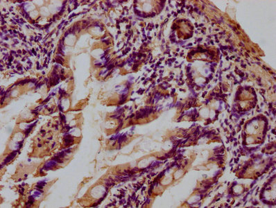
IHC image of CSB-PA012889LA01HU diluted at 1:100 and staining in paraffin-embedded human small intestine tissue performed on a Leica BondTM system. After dewaxing and hydration, antigen retrieval was mediated by high pressure in a citrate buffer (pH 6.0). Section was blocked with 10% normal goat serum 30min at RT. Then primary antibody (1% BSA) was incubated at 4°C overnight. The primary is detected by a biotinylated secondary antibody and visualized using an HRP conjugated SP system.
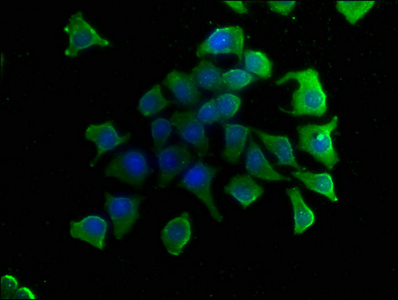
Immunofluorescence staining of A549 cells with CSB-PA012889LA01HU at 1:50, counter-stained with DAPI. The cells were fixed in 4% formaldehyde, permeabilized using 0.2% Triton X-100 and blocked in 10% normal Goat Serum. The cells were then incubated with the antibody overnight at 4°C. The secondary antibody was Alexa Fluor 488-congugated AffiniPure Goat Anti-Rabbit IgG(H+L).
The Rabbit anti-Homo sapiens (Human) LGALS4 Polyclonal antibody is produced in rabbits using Recombinant Human Galectin-4 protein (1-323AA). It is validated for ELISA, IF, and IHC. This antibody reacts with Human Galectin-4 (LGALS4), a protein involved in intracellular and intercellular interactions.
Galectin-4 belongs to a family of proteins that bind to beta-galactoside. It plays a role in cancer and inflammatory diseases of the intestine.
The levels of Galectin-4 in the body have been shown to rise as cancer progresses, making it a possible tumor marker. In inflammatory diseases of the intestine, Galectin-4 stimulates the production of mediators that worsen inflammation.
The Rabbit anti-Homo sapiens (Human) LGALS4 Polyclonal Antibody can be used in research to detect the presence of Galectin-4, a possible tumor marker.


Email: support@cusabio.com
Distributors Worldwide