KLRK1 Antibody

Code: CSB-PA012474LA01HU
Size:
50μl
50μl100μl
US$166
Quantity:
Species Reactivity: Human
Raised in: Rabbit
Application: ELISA, WB, IHC, IF
For inquiries on large quantities or another requirements
Send an Inquiry
Start an on-line Chat
Online ordering is currently available for U.S. customers only. For orders outside the U.S., please kindly submit an inquiry or start a chat with us.

Product Details

Uniprot NO.
Target Names
KLRK1
Alternative Names
KLRK1; D12S2489E; NKG2D; NKG2-D type II integral membrane protein; Killer cell lectin-like receptor subfamily K member 1; NK cell receptor D; NKG2-D-activating NK receptor; CD antigen CD314
Raised in
Rabbit
Species Reactivity
Human
Immunogen
Synthetic peptide of human KLRK1
Immunogen Species
Homo sapiens (Human)
Conjugate
Non-conjugated
Isotype
IgG
Purification Method
Antigen affinity purification
Concentration
It differs from different batches. Please contact us to confirm it.
Buffer
pH7.4 PBS, 0.05% NaN3, 40% Glycerol
Tested Applications
ELISA, WB, IHC, IF
Recommended Dilution
ApplicationRecommended Dilution
WB1:500-1:2000
IHC1:50-1:200
IF1:20-1:100
Storage
Upon receipt, store at -20°C or -80°C. Avoid repeated freeze.
Lead Time
Basically, we can dispatch the products out in 1-3 working days after receiving your orders. Delivery time maybe differs from different purchasing way or location, please kindly consult your local distributors for specific delivery time.
Note: All of our proteins are default shipped with normal blue ice packs, if you request to ship with dry ice, please communicate with us in advance and extra fees will be charged.
Usage
For Research Use Only. Not for use in diagnostic or therapeutic procedures.
Datasheet & COA
Images
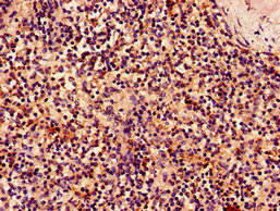
  • IHC image of CSB-PA012474LA01HU diluted at 1:50 and staining in paraffin-embedded human tonsil tissue performed on a Leica BondTM system. After dewaxing and hydration, antigen retrieval was mediated by high pressure in a citrate buffer (pH 6.0). Section was blocked with 10% normal goat serum 30min at RT. Then primary antibody (1% BSA) was incubated at 4°C overnight. The primary is detected by a Goat anti-rabbit polymer IgG labeled by HRP and visualized using 0.05% DAB. Secondary antibody only control: uses 1% BSA instead of primary antibody
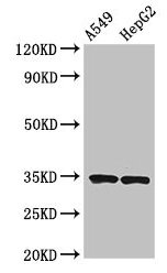
  • Western Blot
    Positive WB detected in: THP-1 whole cell lysate(30µg), A549 whole cell lysate(30µg), U251 whole cell lysate(30µg), Hela whole cell lysate(30µg)
    All lanes: KLRK1 antibody at 1:1000
    Secondary
    Goat polyclonal to rabbit IgG at 1/20000 dilution
    Predicted band size: 26kDa
    Observed band size: 36 kDa
    Exposure time: 120s
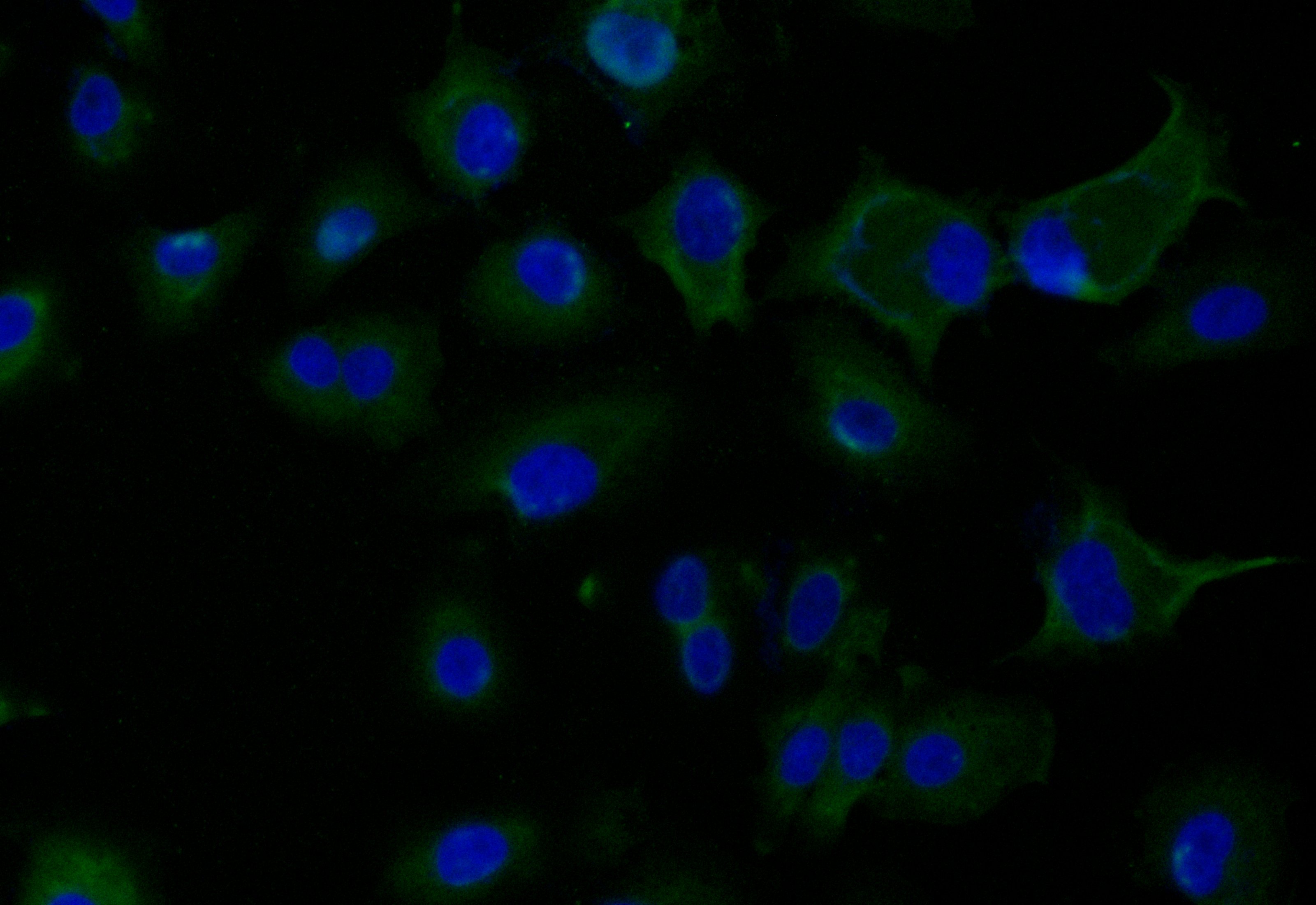
  • Immunofluorescence staining of Hela cell with CSB-PA012474LA01HU at 1:20, counter-stained with DAPI. The cells were fixed in 4% formaldehyde and blocked in 10% normal Goat Serum. The cells were then incubated with the antibody overnight at 4C. The secondary antibody was Alexa Fluor 488-congugated AffiniPure Goat Anti-Rabbit IgG(H+L).
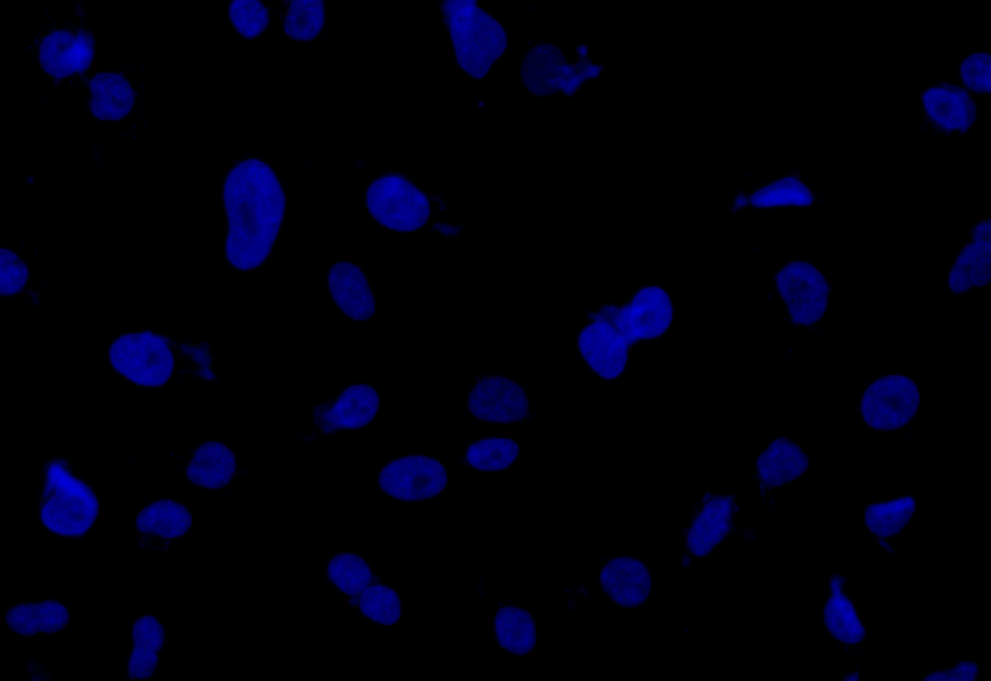
  • Immunofluorescence staining of Hela cell with 5% goat serum, counter-stained with DAPI. The cells were fixed in 4% formaldehyde and blocked in 10% normal Goat Serum. The cells were then incubated with the antibody overnight at 4C. The secondary antibody was Alexa Fluor 488-congugated AffiniPure Goat Anti-Rabbit IgG(H+L).
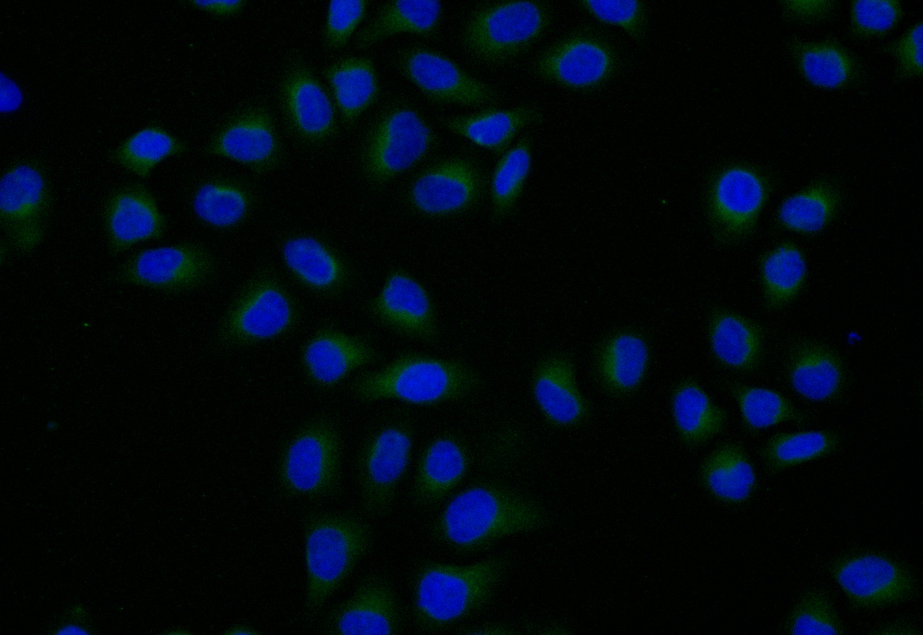
  • Immunofluorescence staining of A549 cell with CSB-PA012474LA01HU at 1:20, counter-stained with DAPI. The cells were fixed in 4% formaldehyde and blocked in 10% normal Goat Serum. The cells were then incubated with the antibody overnight at 4C. The secondary antibody was Alexa Fluor 488-congugated AffiniPure Goat Anti-Rabbit IgG(H+L).
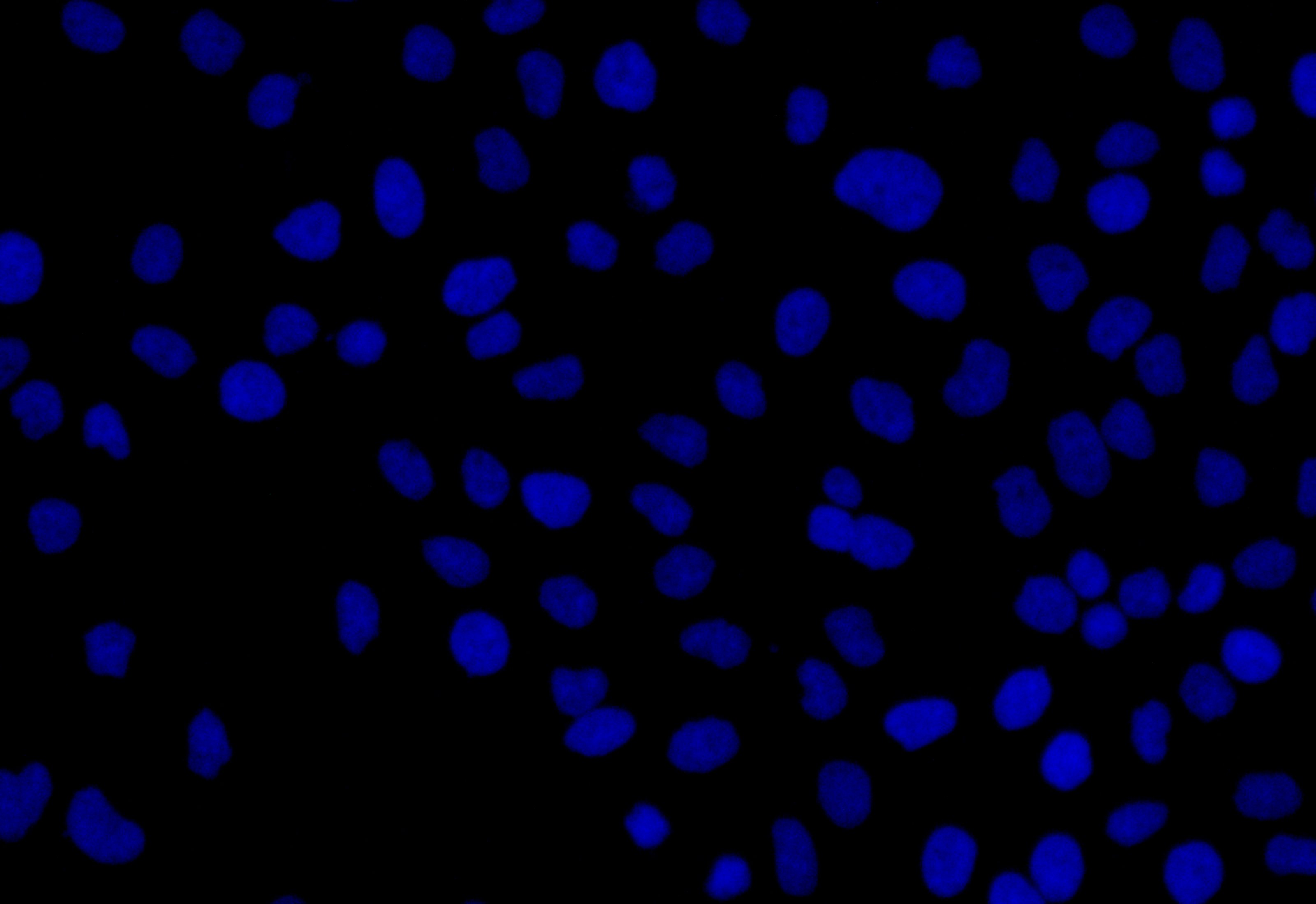
  • Immunofluorescence staining of A549 cell with 5% goat serum, counter-stained with DAPI. The cells were fixed in 4% formaldehyde and blocked in 10% normal Goat Serum. The cells were then incubated with the antibody overnight at 4C. The secondary antibody was Alexa Fluor 488-congugated AffiniPure Goat Anti-Rabbit IgG(H+L).
Description

The KLRK1 polyclonal antibody is a type of antibody that recognizes and binds to the human KLRK1 protein. The KLRK1 protein, also known as NKG2D, mainly functions to recognize and binds to stress-induced ligands expressed on the surface of infected or transformed cells, which allows the immune system to detect and eliminate these cells. KLRK1-mediated cytotoxicity is important for the immune response against viral infections and cancer, as well as for the elimination of cells undergoing cellular stress, such as cells undergoing DNA damage or apoptosis.

KLRK1 antibody is derived from multiple clones of B cells within the rabbit immunized with the recombinant human NKG2D protein (73-216aa), which produces a mixture of antibodies that can recognize different epitopes of the KLRK1 protein. The KLRK1 antibody underwent protein G affinity chromatography, getting a purity exceeding 95%. It has been quality validated in ELISA, WB, IHC, and IF applications.

Customer Reviews and Q&A

 Customer Reviews

Target Background

Function(From Uniprot)
Functions as an activating and costimulatory receptor involved in immunosurveillance upon binding to various cellular stress-inducible ligands displayed at the surface of autologous tumor cells and virus-infected cells. Provides both stimulatory and costimulatory innate immune responses on activated killer (NK) cells, leading to cytotoxic activity. Acts as a costimulatory receptor for T-cell receptor (TCR) in CD8(+) T-cell-mediated adaptive immune responses by amplifying T-cell activation. Stimulates perforin-mediated elimination of ligand-expressing tumor cells. Signaling involves calcium influx, culminating in the expression of TNF-alpha. Participates in NK cell-mediated bone marrow graft rejection. May play a regulatory role in differentiation and survival of NK cells. Binds to ligands belonging to various subfamilies of MHC class I-related glycoproteins including MICA, MICB, RAET1E, RAET1G, RAET1L/ULBP6, ULBP1, ULBP2, ULBP3 (ULBP2>ULBP1>ULBP3) and ULBP4.
Subcellular Location
Cell membrane; Single-pass type II membrane protein. Note=Colocalized with HCST on the cell surface.
Tissue Specificity
Expressed in natural killer (NK) cells, CD8(+) alpha-beta and gamma-delta T-cells. Expressed on essentially all CD56+CD3- NK cells from freshly isolated PBMC. Expressed in interferon-producing killer dendritic cells (IKDCs).
Database Links

HGNC: 18788

UNIGENE: Hs.387787

KEGG: hsa:100528032

STRING: 9606.ENSP00000240618

OMIM: 611817

icon of phone
Call us
301-363-4651 (Available 9 a.m. to 5 p.m. CST from Monday to Friday)

icon of address
Address
No. 269, Shendun 5th Road, Donghu Hi-Tech Development Area, Hubei Province, 430206, P.R.China
icon of social media
Join us with

Subscribe newsletter

Leave a message

* To protect against spam, please pass the CAPTCHA test below.
CAPTCHA verification
© 2007-2025 CUSABIO TECHNOLOGY LLC All rights reserved. 鄂ICP备15011166号-1
Select 0 Products