Call us
301-363-4651 (Available 9 a.m. to 5 p.m. CST from Monday to Friday)
| Application | Recommended Dilution |
|---|---|
| WB | 1:1000-1:5000 |
| IHC | 1:20-1:200 |
| IF | 1:50-1:500 |
| IP | 1:200-1:2000 |
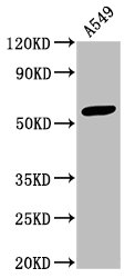
Western Blot
Positive WB detected in: A549 whole cell lysate
All lanes: IFIT3 antibody at 3.3µg/ml
Secondary
Goat polyclonal to rabbit IgG at 1/50000 dilution
Predicted band size: 56 kDa
Observed band size: 56 kDa
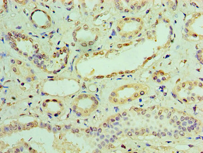
Immunohistochemistry of paraffin-embedded human kidney tissue using CSB-PA011022HA01HU at dilution of 1:100
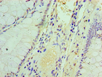
Immunohistochemistry of paraffin-embedded human colon cancer using CSB-PA011022HA01HU at dilution of 1:100
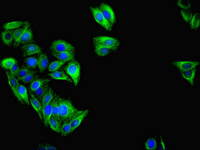
Immunofluorescent analysis of HepG2 cells using CSB-PA011022HA01HU at dilution of 1:100 and Alexa Fluor 488-congugated AffiniPure Goat Anti-Rabbit IgG(H+L)
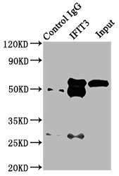
Immunoprecipitating IFIT3 in HepG2 whole cell lysate
Lane 1: Rabbit control IgG (1µg) instead of CSB-PA011022HA01HU in HepG2 whole cell lysate. For western blotting, a HRP-conjugated Protein G antibody was used as the secondary antibody (1/2000)
Lane 2: CSB-PA011022HA01HU (6µg) + HepG2 whole cell lysate (500µg)
Lane 3: HepG2 whole cell lysate (10µg)
To prepare the IFIT3 polyclonal antibody, a rabbit was inoculated with recombinant human 5-hydroxytryptamine receptor 1E protein (203-291aa). Antibodies were extracted from the rabbit serum and then subjected to protein G affinity chromatography purification, resulting in a highly pure antibody with over 95% purity. High purity ensures minimal impurities in this antibody and guarantees its excellent quality.
This IFIT3 polyclonal antibody can effectively recognize and bind to IFIT3 protein from human samples. Its use has been validated in various experimental settings, including ELISA, WB, IHC, IF, and IP. Researchers can employ this antibody to quantitatively or qualitatively measure IFIT3 protein, identify its presence and size, analyze its distribution, and isolate IFIT3 from a mixture solution.
Applications :
Sample type:
Review:
By Anonymous


Email: support@cusabio.com
Distributors Worldwide