
Call us
301-363-4651 (Available 9 a.m. to 5 p.m. CST from Monday to Friday)

| Application | Recommended Dilution |
|---|---|
| WB | 1:500-1:5000 |
| IHC | 1:200-1:500 |
| IF | 1:50-1:200 |
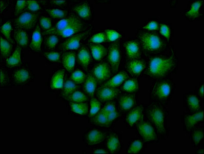
Immunofluorescence staining of Hela cells with CSB-PA009965HA01HU at 1:100, counter-stained with DAPI. The cells were fixed in 4% formaldehyde, permeabilized using 0.2% Triton X-100 and blocked in 10% normal Goat Serum. The cells were then incubated with the antibody overnight at 4°C. The secondary antibody was Alexa Fluor 488-congugated AffiniPure Goat Anti-Rabbit IgG(H+L).
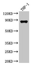
Western Blot
Positive WB detected in: THP-1 whole cell lysate
All lanes: GSN antibody at 3.5µg/ml
Secondary
Goat polyclonal to rabbit IgG at 1/50000 dilution
Predicted band size: 86, 81, 82 kDa
Observed band size: 86 kDa
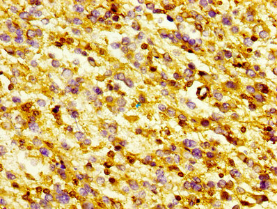
IHC image of CSB-PA009965HA01HU diluted at 1:300 and staining in paraffin-embedded human glioma performed on a Leica BondTM system. After dewaxing and hydration, antigen retrieval was mediated by high pressure in a citrate buffer (pH 6.0). Section was blocked with 10% normal goat serum 30min at RT. Then primary antibody (1% BSA) was incubated at 4°C overnight. The primary is detected by a biotinylated secondary antibody and visualized using an HRP conjugated SP system.
To generate the GSN polyclonal antibody, a rabbit got inoculation with recombinant human Gelsolin protein (28-782aa). Extracting the resulting antibodies from the rabbit serum and then purifying and isolating the GSN polyclonal antibody using protein G affinity chromatography.
This GSN polyclonal antibody has minimal impurities, with a high purity of exceeding 95%. It can recognize and bind to human-origin Gelsolin protein. Additionally, its effectiveness has been validated in a range of experimental settings, including ELISA, WB, IHC, and IF, enabling the detection of levels, determination of presence and size, and analysis of the distribution of the Gelsolin protein.


Email: support@cusabio.com
Distributors Worldwide



