Call us
301-363-4651 (Available 9 a.m. to 5 p.m. CST from Monday to Friday)
| Application | Recommended Dilution |
|---|---|
| IHC | 1:200-1:500 |
| IF | 1:50-1:200 |
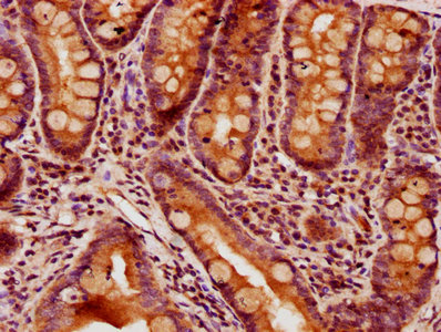
IHC image of CSB-PA885717LA01HU diluted at 1:400 and staining in paraffin-embedded human small intestine tissue performed on a Leica BondTM system. After dewaxing and hydration, antigen retrieval was mediated by high pressure in a citrate buffer (pH 6.0). Section was blocked with 10% normal goat serum 30min at RT. Then primary antibody (1% BSA) was incubated at 4°C overnight. The primary is detected by a biotinylated secondary antibody and visualized using an HRP conjugated SP system.
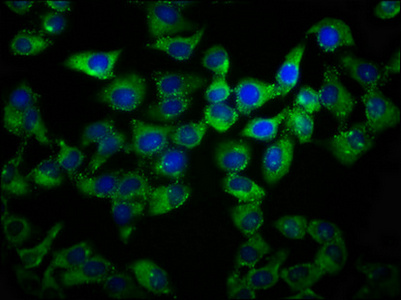
Immunofluorescence staining of Hela cells with CSB-PA885717LA01HU at 1:133, counter-stained with DAPI. The cells were fixed in 4% formaldehyde, permeabilized using 0.2% Triton X-100 and blocked in 10% normal Goat Serum. The cells were then incubated with the antibody overnight at 4°C. The secondary antibody was Alexa Fluor 488-congugated AffiniPure Goat Anti-Rabbit IgG(H+L).


Email: support@cusabio.com
Distributors Worldwide