
Call us
301-363-4651 (Available 9 a.m. to 5 p.m. CST from Monday to Friday)

| Application | Recommended Dilution |
|---|---|
| WB | 1:1000-1:5000 |
| IHC | 1:500-1:1000 |
| IF | 1:200-1:500 |
| IP | 1:200-1:2000 |
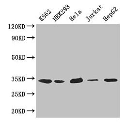
Western Blot
Positive WB detected in: K562 whole cell lysate, HEK293 whole cell lysate, Hela whole cell lysate, Jurkat whole cell lysate, HepG2 whole cell lysate
All lanes: CDK2 antibody at 2.6µg/ml
Secondary
Goat polyclonal to rabbit IgG at 1/50000 dilution
Predicted band size: 34, 31 kDa
Observed band size: 34 kDa
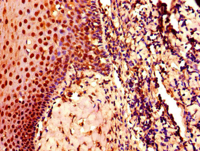
IHC image of CSB-PA005061LA01HU diluted at 1:800 and staining in paraffin-embedded human tonsil tissue performed on a Leica BondTM system. After dewaxing and hydration, antigen retrieval was mediated by high pressure in a citrate buffer (pH 6.0). Section was blocked with 10% normal goat serum 30min at RT. Then primary antibody (1% BSA) was incubated at 4°C overnight. The primary is detected by a biotinylated secondary antibody and visualized using an HRP conjugated SP system.
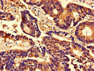
IHC image of CSB-PA005061LA01HU diluted at 1:800 and staining in paraffin-embedded human colon cancer performed on a Leica BondTM system. After dewaxing and hydration, antigen retrieval was mediated by high pressure in a citrate buffer (pH 6.0). Section was blocked with 10% normal goat serum 30min at RT. Then primary antibody (1% BSA) was incubated at 4°C overnight. The primary is detected by a biotinylated secondary antibody and visualized using an HRP conjugated SP system.
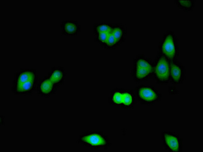
Immunofluorescence staining of HepG2 cells with CSB-PA005061LA01HU at 1:266, counter-stained with DAPI. The cells were fixed in 4% formaldehyde, permeabilized using 0.2% Triton X-100 and blocked in 10% normal Goat Serum. The cells were then incubated with the antibody overnight at 4°C. The secondary antibody was Alexa Fluor 488-congugated AffiniPure Goat Anti-Rabbit IgG(H+L).
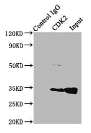
Immunoprecipitating CDK2 in Hela whole cell lysate
Lane 1: Rabbit control IgG (1µg) instead of CSB-PA005061LA01HU in Hela whole cell lysate. For western blotting, a HRP-conjugated Protein G antibody was used as the secondary antibody (1/50000)
Lane 2: CSB-PA005061LA01HU (4µg) + Hela whole cell lysate (500µg)
Lane 3: Hela whole cell lysate (20µg)
The CDK2 polyclonal antibody is raised in rabbits and specifically recognizes and binds to the CDK2 protein. The immunogen used to generate this antibody is the recombinant human CDK2 protein consisting of 1-298 amino acids. After immunization, the antibody was collected, isolated, and purified through protein G chromatography, which results in a purity level of >95%.
This antibody can react with human samples, and it has been extensively tested in various applications such as ELISA, WB, IHC, IF, and IP. Therefore, the CDK2 polyclonal antibody can be used to investigate the expression, localization, and activity of CDK2 in various experimental contexts, aiding in the understanding of its roles in normal and disease states.


Email: support@cusabio.com
Distributors Worldwide



