Call us
301-363-4651 (Available 9 a.m. to 5 p.m. CST from Monday to Friday)
| Application | Recommended Dilution |
|---|---|
| WB | 1:100-1:1000 |
| ICC | 1:20-1:200 |
| IF | 1:20-1:200 |
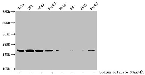
Western Blot
Detected samples: Hela whole cell lysate, 293 whole cell lysate, A549 whole cell lysate, HepG2 whole cell lysate; Untreated (-) or treated (+) with 30mM sodium butyrate for 4h
All lanes: HIST1H1C antibody at 3.5µg/ml
Secondary
Goat polyclonal to rabbit IgG at 1/50000 dilution
Predicted band size: 22 kDa
Observed band size: 22 kDa
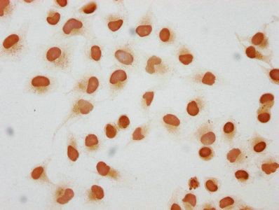
Immunocytochemistry analysis of CSB-PA010378OA109hibHU diluted at 1:50 and staining in Hela cells (treated with 30mM sodium butyrate for 4h) performed on a Leica BondTM system. The cells were fixed in 4% formaldehyde, permeabilized using 0.2% Triton X-100 and blocked with 10% normal goat serum 30min at RT. Then primary antibody (1% BSA) was incubated at 4°C overnight. The primary is detected by a biotinylated secondary antibody and visualized using an HRP conjugated SP system.
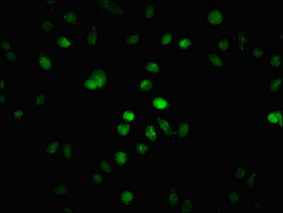
Immunofluorescence staining of Hela cells (treated with 30mM sodium butyrate for 4h) with CSB-PA010378OA109hibHU at 1:25, counter-stained with DAPI. The cells were fixed in 4% formaldehyde, permeabilized using 0.2% Triton X-100 and blocked in 10% normal Goat Serum. The cells were then incubated with the antibody overnight at 4°C. The secondary antibody was Alexa Fluor 488-congugated AffiniPure Goat Anti-Rabbit IgG(H+L).
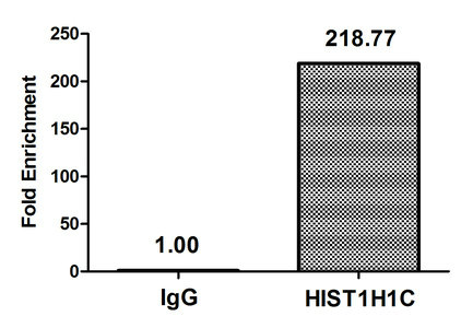
Chromatin Immunoprecipitation Hela (4*106, treated with 30mM sodium butyrate for 4h) were treated with Micrococcal Nuclease, sonicated, and immunoprecipitated with 5µg anti-HIST1H1C (CSB-PA010378OA109hibHU) or a control normal rabbit IgG. The resulting ChIP DNA was quantified using real-time PCR with primers against the β-Globin promoter.
The 2-hydroxyisobutyryl-HIST1H1C (K109) Antibody was raised in a rabbit using a peptide around the site of 2-hydroxyisobutyryl-Lys (109) derived from Human Histone H1.2 as the immunogen. It’s a polyclonal, non-conjugated, IgG purified by antigen affinity. It finds uses in ELISA, Western blot (WB), Immunocytochemistry (ICC), Immunofluorescence (IF), and Chromatin Immunoprecipitation (ChIP). This antibody reacts against the human histone HIST1H1C. It can detect the endogenous levels of HIST1H1C of human-origin. At the moment, there’s no function assigned to the K109 2-hydroxyisobutyryl modified lysine, and it’s still in need to be dilucidated. The HIST1H1C protein interacts with the linker DNA between nucleosomes, facilitating the chromatin condensation to higher-order fibers. And this histone also can affect the nucleosome spacing, chromatin remodeling, and DNA methylation, consequently modulating the gene expression. Therefore, it is vital for correct chromatin high structure formation, regulation, and maintenance.


Email: support@cusabio.com
Distributors Worldwide