
Call us
301-363-4651 (Available 9 a.m. to 5 p.m. CST from Monday to Friday)

| Application | Recommended Dilution |
|---|---|
| WB | 1:1000-1:5000 |
| IHC | 1:50-1:200 |
| IF | 1:50-1:200 |
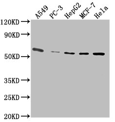
Western Blot
Positive WB detected in: A549 whole cell lysate, PC-3 whole cell lysate, HepG2 whole cell lysate, MCF-7 whole cell lysate, Hela whole cell lysate
All lanes: PD-L1 antibody at 1:2500
Secondary
Goat polyclonal to Mouse IgG at 1/10000 dilution
Predicted band size: 34, 21 kDa
Observed band size: 55 kDa
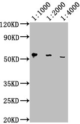
Western Blot
Positive WB detected in: Hela whole cell lysate
All lanes: PD-L1 antibody at 1:1000, 1:2000, 1:4000
Secondary
Goat polyclonal to Mouse IgG at 1/10000 dilution
Predicted band size: 34, 21 kDa
Observed band size: 55 kDa
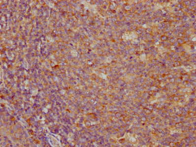
IHC image of CSB-MA878942A0m diluted at 1:100 and staining in paraffin-embedded human tonsil tissue performed on a Leica BondTM system. After dewaxing and hydration, antigen retrieval was mediated by high pressure in a citrate buffer (pH 6.0). Section was blocked with 10% normal goat serum 30min at RT. Then primary antibody (1% BSA) was incubated at 4°C overnight. The primary is detected by a biotinylated secondary antibody and visualized using an HRP conjugated SP system.
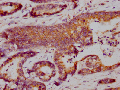
IHC image of CSB-MA878942A0m diluted at 1:100 and staining in paraffin-embedded human colon cancer performed on a Leica BondTM system. After dewaxing and hydration, antigen retrieval was mediated by high pressure in a citrate buffer (pH 6.0). Section was blocked with 10% normal goat serum 30min at RT. Then primary antibody (1% BSA) was incubated at 4°C overnight. The primary is detected by a biotinylated secondary antibody and visualized using an HRP conjugated SP system.
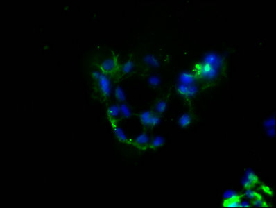
Immunofluorescence staining of 293 cells with CSB-MA878942A0m at 1:100, counter-stained with DAPI. The cells were blocked in 10% normal Goat Serum and then incubated with the primary antibody overnight at 4°C. The secondary antibody was Alexa Fluor 488-congugated AffiniPure Goat Anti-Mouse IgG(H+L).
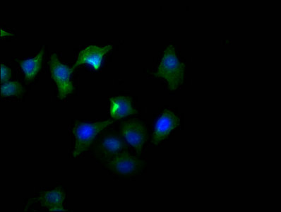
Immunofluorescence staining of A549 cells with CSB-MA878942A0m at 1:100, counter-stained with DAPI. The cells were blocked in 10% normal Goat Serum and then incubated with the primary antibody overnight at 4°C. The secondary antibody was Alexa Fluor 488-congugated AffiniPure Goat Anti-Mouse IgG(H+L).
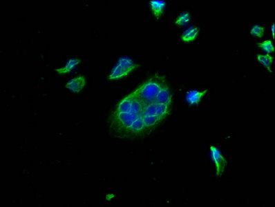
Immunofluorescence staining of Hela cells with CSB-MA878942A0m at 1:100, counter-stained with DAPI. The cells were blocked in 10% normal Goat Serum and then incubated with the primary antibody overnight at 4°C. The secondary antibody was Alexa Fluor 488-congugated AffiniPure Goat Anti-Mouse IgG(H+L).
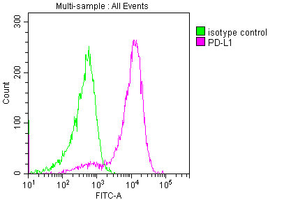
Overlay histogram showing 293 cells stained with CSB-MA878942A0m (red line) at 1:150. The cells were incubated in 1x PBS /10% normal goat serum to block non-specific protein-protein interactions followed by primary antibody for 1 h at 4°C. The secondary antibody used was FITC goat anti-mouse IgG(H+L) at 1/200 dilution for 1 h at 4°C. Isotype control antibody (green line) was used under the same conditions. Acquisition of >10,000 events was performed.
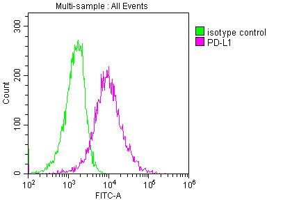
Overlay histogram showing A549 cells stained with CSB-MA878942A0m (red line) at 1:150. The cells were incubated in 1x PBS /10% normal goat serum to block non-specific protein-protein interactions followed by primary antibody for 1 h at 4°C. The secondary antibody used was FITC goat anti-mouse IgG(H+L) at 1/200 dilution for 1 h at 4°C. Isotype control antibody (green line) was used under the same conditions. Acquisition of >10,000 events was performed.
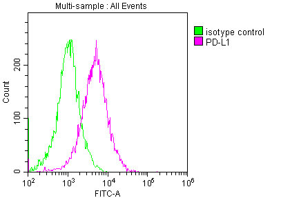
Overlay histogram showing Hela cells stained with CSB-MA878942A0m (red line) at 1:150. The cells were incubated in 1x PBS /10% normal goat serum to block non-specific protein-protein interactions followed by primary antibody for 1 h at 4°C. The secondary antibody used was FITC goat anti-mouse IgG(H+L) at 1/200 dilution for 1 h at 4°C. Isotype control antibody (green line) was used under the same conditions. Acquisition of >10,000 events was performed.
The PD-L1 monoclonal antibody was harvested from the hybridomas, which were formed by fusing the spleen cells and myeloma cells. The spleen cells were isolated from the mouse immunized with recombinant human PD-L1 protein (19-238aa). The resulting PD-L1 monoclonal antibody is highly specific for human PD-L1 protein and can be used in a variety of research applications, including ELISA, WB, IHC, IF, and FC. Protein G purification of this antibody brought its purity up to 95%.
PD-L1 mainly regulates the immune system by interacting with its receptor PD-1, which is found on activated T cells. The binding of PD-L1 to PD-1 leads to the inhibition of T-cell activity, preventing excessive immune responses that can result in tissue damage and autoimmune disorders. PD-L1 is also exploited by cancer cells as a mechanism of immune evasion, allowing them to evade attack by the immune system.
Applications :
Sample type:
Sample dilution:
Review:
By Anonymous


Email: support@cusabio.com
Distributors Worldwide



