Call us
301-363-4651 (Available 9 a.m. to 5 p.m. CST from Monday to Friday)
| Application | Recommended Dilution |
|---|---|
| WB | 1:500-1:5000 |
| IHC | 1:50-1:500 |
| IF | 1:50-1:200 |
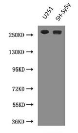
Western Blot
Positive WB detected in: U251 whole cell lysate, SH-SY5Y whole cell lysate
All lanes: NES antibody at 3µg/ml
Secondary
Goat polyclonal to Mouse IgG at 1/10000 dilution
Predicted band size: 178 kDa
Observed band size: 260 kDa
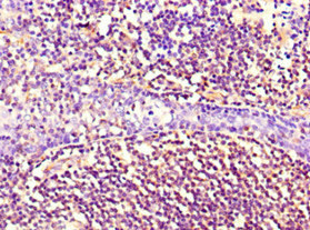
Immunohistochemistry of paraffin-embedded human tonsil tissue using CSB-MA0157131A0m at dilution of 1:100
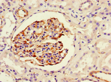
Immunohistochemistry of paraffin-embedded human kidney tissue using CSB-MA0157131A0m at dilution of 1:100
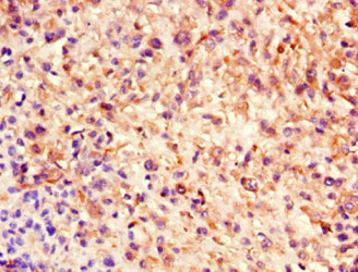
Immunohistochemistry of paraffin-embedded human melanoma using CSB-MA0157131A0m at dilution of 1:100
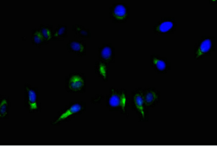
Immunofluorescent analysis of Hela cells using CSB-MA0157131A0m at a dilution of 1:100 and Alexa Fluor 488-congugated AffiniPure Goat Anti-Mouse IgG(H+L).
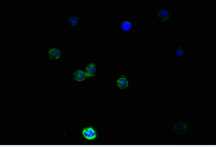
Immunofluorescent analysis of PC-3 cells using CSB-MA0157131A0m at a dilution of 1:100 and Alexa Fluor 488-congugated AffiniPure Goat Anti-Mouse IgG(H+L).
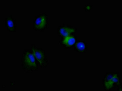
Immunofluorescent analysis of U251 cells using CSB-MA0157131A0m at a dilution of 1:100 and Alexa Fluor 488-congugated AffiniPure Goat Anti-Mouse IgG(H+L).
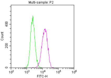
Overlay histogram showing U251 cells stained with CSB-MA0157131A0m (red line). The cells were fixed with 70% Ethylalcohol (18h) and then permeabilized with 0.3% Triton X-100 for 2 min. The cells were then incubated in 1x PBS /10% normal goat serum to block non-specific protein-protein interactions followed by the antibody (10µg/1*106cells) for 1 h at 4°C. The secondary antibody used was FITC goat anti-mouse IgG(H+L) at 1/200 dilution for 1 h at 4°C. Isotype control antibody (green line) was mouse IgG1 (10µg/1*106cells) used under the same conditions. Acquisition of >10,000 events was performed.
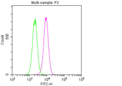
Overlay histogram showing Hela cells stained with CSB-MA0157131A0m (red line). The cells were fixed with 70% Ethylalcohol (18h) and then permeabilized with 0.3% Triton X-100 for 2 min. The cells were then incubated in 1x PBS /10% normal goat serum to block non-specific protein-protein interactions followed by the antibody (10µg/1*106cells) for 1 h at 4°C. The secondary antibody used was FITC goat anti-mouse IgG(H+L) at 1/200 dilution for 1 h at 4°C. Isotype control antibody (green line) was mouse IgG1 (10µg/1*106cells) used under the same conditions. Acquisition of >10,000 events was performed.
This (Nestin) NES antibody is a monoclonal antibody matched with the isotype IgG1. It is generated through sequential procedures, including the immunization of mice to get NES-producing B cells, the fusion of NES-producing B cells with myeloma cells, the screening of hybridoma cells, and the inoculation of the hybridoma cells into mice. Its immunogen is the recombinant human NES protein. The mouse ascites collected underwent protein G-mediated purification, finally harvesting the NES antibody with 95%+ purity. This NES monoclonal antibody is recommended for use in ELISA, WB, IHC, IF, and FC analyses. And it only shows reactivity with the samples containing human NES protein.
Nestin, a type VI intermediate filament, constitutes a major component of the cytoskeleton. It may participate in the modulation of assembly and disassembly of vimentin during mitosis. And it also mediates the interaction between intermediate filaments with microtubules and/or microfilaments. Importantly, nestin plays a role in essential stem cell functions, including self-renewal/proliferation, differentiation, and migration, in the context of the cytoskeleton.


Email: support@cusabio.com
Distributors Worldwide