Call us
301-363-4651 (Available 9 a.m. to 5 p.m. CST from Monday to Friday)
| Application | Recommended Dilution |
|---|---|
| WB | 1:500-1:5000 |
| IHC | 1:50-1:500 |
| IF | 1:50-1:200 |
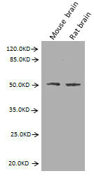
Western Blot
Positive WB detected in: Mouse brain tissue, Rat brain tissue
All lanes: GFAP antibody at 2.7µg/ml
Secondary
Goat polyclonal to Mouse IgG at 1/10000 dilution
Predicted band size: 50, 51 kDa
Observed band size: 50 kDa
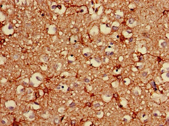
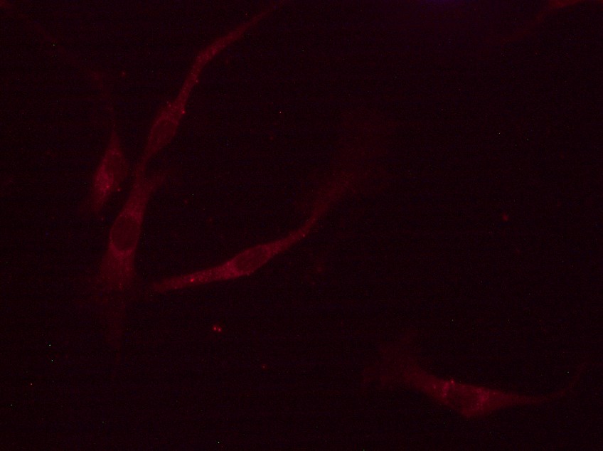
Immunohistochemistry of paraffin-embedded human brain tissue using CSB-MA009369A0m at dilution of 1:100
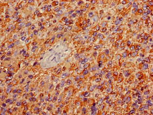
Immunohistochemistry of paraffin-embedded human glioma using CSB-MA009369A0m at dilution of 1:100
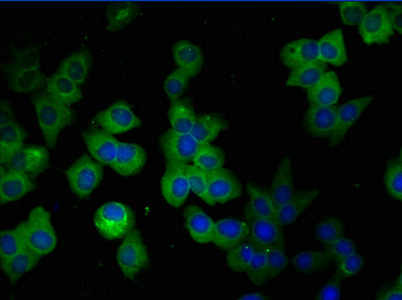
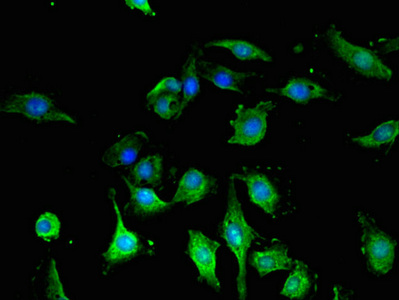
Immunofluorescent analysis of SH-SY5Y cells using CSB-MA009369A0m at a dilution of 1:100 and Alexa Fluor 488-congugated AffiniPure Goat Anti-Mouse IgG(H+L).
Immunofluorescent analysis of U251 cells using CSB-MA009369A0m at a dilution of 1:100 and Alexa Fluor 488-congugated AffiniPure Goat Anti-Mouse IgG(H+L).
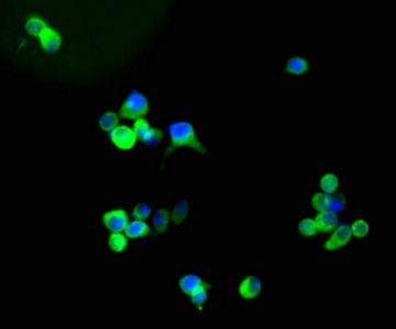
Immunofluorescent analysis of U87 cells using CSB-MA009369A0m at a dilution of 1:100 and Alexa Fluor 488-congugated AffiniPure Goat Anti-Mouse IgG(H+L).
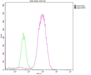
Overlay histogram showing SH-SY5Y cells stained with CSB-MA009369A0m (red line). The cells were fixed with 70% Ethylalcohol (18h) and then permeabilized with 0.3% Triton X-100 for 2 min. The cells were then incubated in 1x PBS /10% normal goat serum to block non-specific protein-protein interactions followed by the antibody (10µg/1*106cells) for 1 h at 4°C. The secondary antibody used was FITC goat anti-mouse IgG(H+L) at 1/200 dilution for 1 h at 4°C. Isotype control antibody (green line) was mouse IgG2b (10µg/1*106cells) used under the same conditions. Acquisition of >10,000 events was performed.
The product CSB-MA009369A0m is an unconjugated monoclonal antibody against glial fibrillary acidic protein (GFAP) from human. It is derived from the mouse myeloma cell-splenocyte hybridoma. The splenocyte is screened from the mouse immunized with the recombinant human GFAP protein (292-432aa) and can secret GFAP antibodies. This GFAP antibody can react with GFAP protein from human, mouse, and rat. It is matched with the mouse IgG2b isotype. Protein G-mediated purification of this GFAP antibody makes its purity up to more than 95%. This antibody has been validated for use in ELISA, WB, IHC, IF, and FC applications.
GFAP is an intermediate filament expressed primarily by astrocytes in the central nervous system (CNS). It mainly maintains astrocyte structural integrity and helps cell movement and shape change. GFAP'sinteraction with glutamate aspartate transporter (GLAST) influences astrocytic functions such as glutamate homeostasis.
Applications :
Sample type:
Sample dilution:
Review:
By Anonymous


Email: support@cusabio.com
Distributors Worldwide