Call us
301-363-4651 (Available 9 a.m. to 5 p.m. CST from Monday to Friday)
| Application | Recommended Dilution |
|---|---|
| WB | 1:2000-1:32000 |
| IHC | 1:200-1:600 |
| IF | 1:100-1:300 |
| FC | 1:200-1:600 |
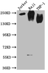
Western Blot
Positive WB detected in: Jurkat whole cell lysate, Raji whole cell lysate, THP-1 whole cell lysate
All lanes CD45 antibody at 1:2000
Secondary
Goat polyclonal to mouse IgG at 1/50000 dilution
Predicted band size: 148, 132, 143, 141, 139, 136 KDa
Observed band size: 180-250 KDa
Exposure time:15min
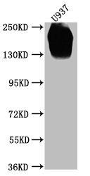
Western Blot
Positive WB detected in: U937 whole cell lysate
All lanes CD45 antibody at 1:2000
Secondary
Goat polyclonal to mouse IgG at 1/50000 dilution
Predicted band size: 148, 132, 143, 141, 139, 136 KDa
Observed band size: 180-250 KDa
Exposure time:5min
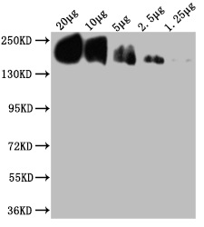
Western Blot
Positive WB detected in: THP-1whole cell lysate at 20μg, 10μg, 5μg, 2.5μg, 1.25μg All lanes: CD45 antibody at 1:2000
Secondary
Goat polyclonal to mouse IgG at 1/50000 dilution
Predicted band size: 148, 132, 143, 141, 139, 136 KDa
Observed band size: 180-250 KDa
Exposure time:15min
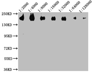
Western Blot
Positive WB detected in: 20μg THP-1 whole cell lysate CD45 antibody at 1:2000, 1:4000, 1:8000, 1:16000, 1:32000, 1:64000, 1:128000
Secondary
Goat polyclonal to mouse IgG at 1/50000 dilution
Predicted band size: 148, 132, 143, 141, 139, 136 KDa
Observed band size: 180-250 KDa
Exposure time:15min
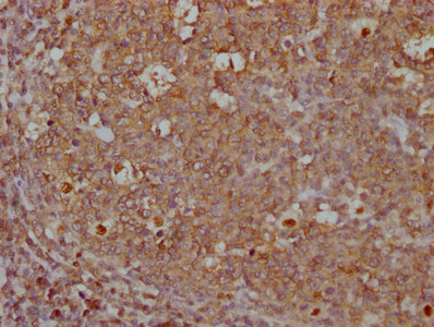
IHC image of CSB-MA019049A0m diluted at 1:500 and staining in paraffin-embedded human tonsil tissue performed on a Leica BondTM system. After dewaxing and hydration, antigen retrieval was mediated by high pressure in a citrate buffer (pH 6.0). Section was blocked with 10% normal goat serum 30min at RT. Then primary antibody (1% BSA) was incubated at 4°C overnight. The primary is detected by a Goat anti-rabbit IgG labeled by HRP and visualized using 0.05% DAB.
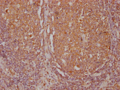
IHC image of CSB-MA019049A0m diluted at 1:500 and staining in paraffin-embedded human lymph node tissue performed on a Leica BondTM system. After dewaxing and hydration, antigen retrieval was mediated by high pressure in a citrate buffer (pH 6.0). Section was blocked with 10% normal goat serum 30min at RT. Then primary antibody (1% BSA) was incubated at 4°C overnight. The primary is detected by a Goat anti-rabbit IgG labeled by HRP and visualized using 0.05% DAB.
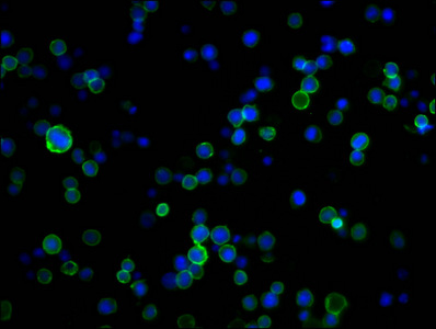
Immunofluorescence staining of Jurkat cells with CSB-MA019049A0m at 1:250, counter-stained with DAPI. The cells were incubated with the antibody overnight at 4°C. Nuclear DNA was labeled in blue with DAPI. The secondary antibody was FITC-conjugated AffiniPure Goat Anti-Mouse IgG (H+L).
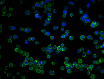
Immunofluorescence staining of Raji cells with CSB-MA019049A0m at 1:250, counter-stained with DAPI. The cells were incubated with the antibody overnight at 4°C. Nuclear DNA was labeled in blue with DAPI. The secondary antibody was FITC-conjugated AffiniPure Goat Anti-Mouse IgG (H+L).
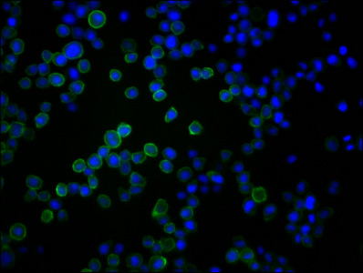
Immunofluorescence staining of U937 cells with CSB-MA019049A0m at 1:250, counter-stained with DAPI. The cells were incubated with the antibody overnight at 4°C. Nuclear DNA was labeled in blue with DAPI. The secondary antibody was FITC-conjugated AffiniPure Goat Anti-Mouse IgG (H+L).
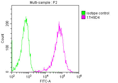
Overlay histogram showing Jurkat cells stained with CSB-MA019049A0m (red line) at 1:500. The cells were incubated in 10% normal goat serum to block non-specific protein-protein interactions followed by the antibody (1µg/1*106cells) for 1 h at 4°C. The secondary antibody used was FITC-conjugated Goat Anti-Mouse IgG(H+L) at 1/100 dilution for 30min at 4°C. Isotype control antibody (green line) was mouse IgG2b (1µg/1*106cells) used under the same conditions. Acquisition of >10,000 events was performed.
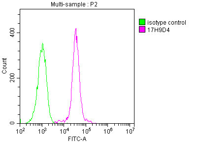
Overlay histogram showing Raji cells stained with CSB-MA019049A0m (red line) at 1:500. The cells were incubated in 10% normal goat serum to block non-specific protein-protein interactions followed by the antibody (1µg/1*106cells) for 1 h at 4°C. The secondary antibody used was FITC-conjugated Goat Anti-Mouse IgG(H+L) at 1/100 dilution for 30min at 4°C. Isotype control antibody (green line) was mouse IgG2b (1µg/1*106cells) used under the same conditions. Acquisition of >10,000 events was performed.
This is a mouse monoclonal CD45 antibody generated by immunizing mice with the recombinant human receptor-type tyrosine-protein phosphatase C protein (24-575AA). Its isotype is IgG2b. This CD45 monoclonal antibody is recommended for ELISA, WB, IHC, IF, and FC applications and can detect CD45 protein of human origin. This monoclonal CD45 antibody is purified using protein A and reaches up to 95% in purity.
The target CD45 protein is an abundant leukocyte cell surface glycoprotein exclusively upon cells of the hematopoietic system. CD45 has been considered as a useful biomarker for the differential diagnosis of undifferentiated lymphoma due to its leukocyte-specific distribution. Alternative splicing makes CD45 protein present multiple isoforms, which share identical transmembrane and cytoplasmic domains but differ in extracellular domains. CD45 glycoprotein plays a crucial role in the antigen-stimulated proliferation of T lymphocytes and thymic development. It is also an essential regulator of T and B cell antigen receptor-mediated activation. As a receptor-type protein tyrosine phosphatase (PTP), CD45 can either positively or negatively regulate Src-family protein tyrosine kinase (PTK) activity in vivo.


Email: support@cusabio.com
Distributors Worldwide