
Call us
301-363-4651 (Available 9 a.m. to 5 p.m. CST from Monday to Friday)

| Application | Recommended Dilution |
|---|---|
| WB | 1:1000-1:40000 |
| IHC | 1:50-1:200 |
| IF | 1:50-1:200 |
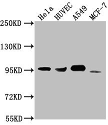
Western Blot
Positive WB detected in: Hela whole cell lysate, HUVEC whole cell lysate, A549 whole cell lysate, MCF-7 whole cell lysate
All lanes: CD44 antibody at 1:1500
Secondary
Goat polyclonal to Mouse IgG at 1/10000 dilution
Predicted band size: 82, 4, 78, 77, 81, 79, 75, 54,47, 40, 44, 33, 74, 76, 38, 16 kDa
Observed band size: 95 kDa
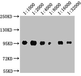
Western Blot
Positive WB detected in: A549 whole cell lysate
All lanes: CD44 antibody at 1:1000, 1:2000, 1:4000, 1:8000, 1:16000, 1:32000
Secondary
Goat polyclonal to Mouse IgG at 1/10000 dilution
Predicted band size: 82, 4, 78, 77, 81, 79, 75, 54,47, 40, 44, 33, 74, 76, 38, 16 kDa
Observed band size: 95 kDa
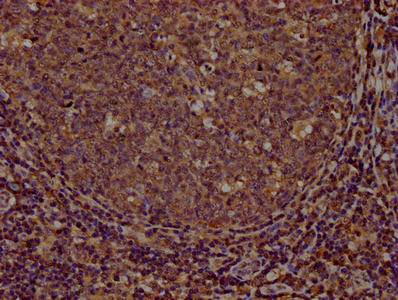
IHC image of CSB-MA004938A0m diluted at 1:100 and staining in paraffin-embedded human tonsil tissue performed on a Leica BondTM system. After dewaxing and hydration, antigen retrieval was mediated by high pressure in a citrate buffer (pH 6.0). Section was blocked with 10% normal goat serum 30min at RT. Then primary antibody (1% BSA) was incubated at 4°C overnight. The primary is detected by a biotinylated secondary antibody and visualized using an HRP conjugated SP system.
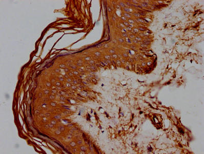
IHC image of CSB-MA004938A0m diluted at 1:100 and staining in paraffin-embedded human skin tissue performed on a Leica BondTM system. After dewaxing and hydration, antigen retrieval was mediated by high pressure in a citrate buffer (pH 6.0). Section was blocked with 10% normal goat serum 30min at RT. Then primary antibody (1% BSA) was incubated at 4°C overnight. The primary is detected by a biotinylated secondary antibody and visualized using an HRP conjugated SP system.
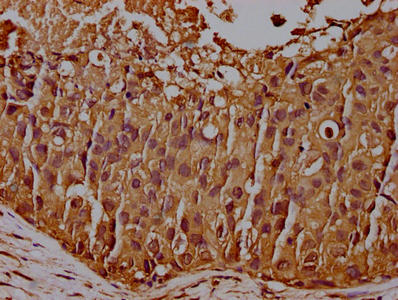
IHC image of CSB-MA004938A0m diluted at 1:100 and staining in paraffin-embedded human breast cancer performed on a Leica BondTM system. After dewaxing and hydration, antigen retrieval was mediated by high pressure in a citrate buffer (pH 6.0). Section was blocked with 10% normal goat serum 30min at RT. Then primary antibody (1% BSA) was incubated at 4°C overnight. The primary is detected by a biotinylated secondary antibody and visualized using an HRP conjugated SP system.
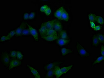
Immunofluorescence staining of Hela cells with CSB-MA004938A0m at 1:100, counter-stained with DAPI. The cells were blocked in 10% normal Goat Serum and then incubated with the primary antibody overnight at 4°C. The secondary antibody was Alexa Fluor 488-congugated AffiniPure Goat Anti-Mouse IgG(H+L).
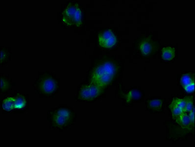
Immunofluorescence staining of MCF-7 cells with CSB-MA004938A0m at 1:100, counter-stained with DAPI. The cells were blocked in 10% normal Goat Serum and then incubated with the primary antibody overnight at 4°C. The secondary antibody was Alexa Fluor 488-congugated AffiniPure Goat Anti-Mouse IgG(H+L).
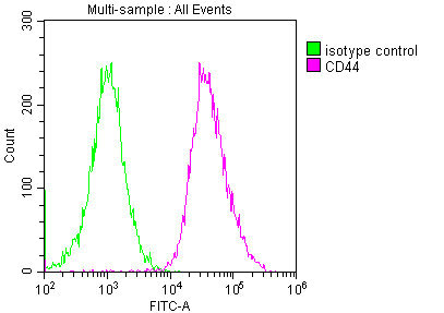
Overlay histogram showing Hela cells stained with CSB-MA004938A0m (red line) at 1:200. The cells were incubated in 1x PBS /10% normal goat serum to block non-specific protein-protein interactions followed by primary antibody for 1 h at 4°C. The secondary antibody used was FITC goat anti-mouse IgG(H+L) at 1/200 dilution for 1 h at 4°C. Isotype control antibody (green line) was used under the same conditions. Acquisition of >10,000 events was performed.
This CD44 monoclonal antibody was produced against a Recombinant Human CD44 antigen protein in order to bind to CD44 antigens. Fusion of cell culture myeloma cells with mouse spleen cells antibodies resulted in hybrid cells which produced large amounts of CD44 monoclonal antibody. The cell fusion resulted in the cells with two properties, one with the ability to grow continually, and the other with the ability to produce the CD44 monoclonal antibody, which has been validated in ELISA, WB, IHC, IF, FC.
CD44 belongs to the family of adhesion factors and is a type of transmembrane single-chain glycoprotein widely expressed on the cell surface. Originally described as a granulocyte-T lymphocyte antigen, it was named the CD44 cluster at the third international working group meeting of leukocyte differentiation antigens. CD44 is a family of glycoproteins encoded by a single gene located on chromosome 11p13. The molecular mass is between 80 and 200 kDa. CD44 is highly glycosylated, including N-linked and O-linked glycosylation and chondroitin sulfate. Heparinized.

Email: support@cusabio.com
Distributors Worldwide



