
Call us
301-363-4651 (Available 9 a.m. to 5 p.m. CST from Monday to Friday)

| Application | Recommended Dilution |
|---|---|
| WB | 1:500-1:2000 |
| IF | 1:50-1:200 |
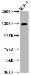
Western Blot
Positive WB detected in: MCF-7 whole cell lysate
All lanes: CD146 antibody at 1:1250
Secondary
Goat polyclonal to Mouse IgG at 1/50000 dilution
Predicted band size: 72, 58 kDa
Observed band size: 120 kDa
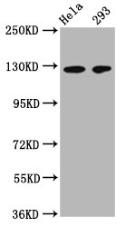
Western Blot
Positive WB detected in: Hela whole cell lysate, 293 whole cell lysate
All lanes: CD146 antibody at 1:500
Secondary
Goat polyclonal to Mouse IgG at 1/50000 dilution
Predicted band size: 72, 58 kDa
Observed band size: 120 kDa
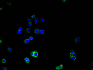
Immunofluorescence staining of A375 cells with CSB-MA013563A0m at 1:100, counter-stained with DAPI. The cells were blocked in 10% normal Goat Serum and then incubated with the primary antibody overnight at 4°C. The secondary antibody was Alexa Fluor 488-congugated AffiniPure Goat Anti-Mouse IgG(H+L).
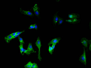
Immunofluorescence staining of Hela cells with CSB-MA013563A0m at 1:100, counter-stained with DAPI. The cells were blocked in 10% normal Goat Serum and then incubated with the primary antibody overnight at 4°C. The secondary antibody was Alexa Fluor 488-congugated AffiniPure Goat Anti-Mouse IgG(H+L).
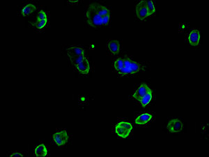
Immunofluorescence staining of MCF-7 cells with CSB-MA013563A0m at 1:100, counter-stained with DAPI. The cells were blocked in 10% normal Goat Serum and then incubated with the primary antibody overnight at 4°C. The secondary antibody was Alexa Fluor 488-congugated AffiniPure Goat Anti-Mouse IgG(H+L).
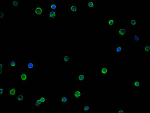
Immunofluorescence staining of THP-1 cells with CSB-MA013563A0m at 1:100, counter-stained with DAPI. The cells were blocked in 10% normal Goat Serum and then incubated with the primary antibody overnight at 4°C. The secondary antibody was Alexa Fluor 488-congugated AffiniPure Goat Anti-Mouse IgG(H+L).
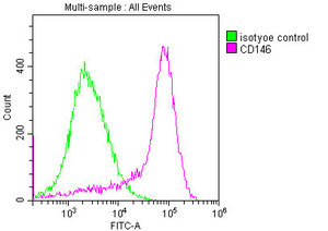
Overlay histogram showing MCF-7 cells stained with CSB-MA013563A0m (red line) at 1:400. The cells were incubated in 1x PBS /10% normal goat serum to block non-specific protein-protein interactions followed by primary antibody for 1 h at 4°C. The secondary antibody used was FITC goat anti-mouse IgG(H+L) at 1/200 dilution for 1 h at 4°C. Isotype control antibody (green line) was used under the same conditions. Acquisition of >10,000 events was performed.
The monoclonal anti-CD146 (mouse IgG2a isotype) is produced from the hybridoma fused by the mouse myeloma cells and splenocytes from the immunized mouse. The splenocytes are isolated from the mouse immunized with recombinant human CD146 protein (50-646AA) and can secret CD146 antibodies. This unconjugated CD146 antibody is purified through protein G with a purity of more than 95%. It only reacts with samples containing CD146 from human. And it has been amenable for ELISA, WB, IF, and FC applications.
The CD146 functions as a Ca2+-independent cell adhesion molecule involved in heterophilic cell-cell interactions. It also participates in inflammation, differentiation, adhesion, tumourigenicity, metastasis, invasion, and angiogenesis.

Applications :
Sample type:
Sample dilution:
Review:
By Anonymous
Applications :
Sample type:
Sample dilution:
Review:
By Anonymous


Email: support@cusabio.com
Distributors Worldwide



