Mouse Vascular cell adhesion molecule 1,VCAM-1 ELISA kit

Promotion
Code: CSB-E04754m
Size:
96T,5×96T,10×96T
Pls inquire
Quantity:
Sample Types: serum, plasma, cell culture supernates, tissue homogenates
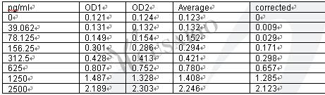
Detect Range: 39.062 pg/mL-2500 pg/mL
Sensitivity: 29.649 pg/mL
Trial Size:
24T ELISA Kit Trial Size (Only USD$150/ kit)
* Sample kit cost can be deducted as a $30 credit for each 96-assay kit of the same analyte and brand you subsequently purchase within six months until depleted. More details >>
Interested in a trial size? Please leave a message below.

For inquiries on large quantities or another requirements
Send an Inquiry
Start an on-line Chat
Online ordering is currently available for U.S. customers only. For orders outside the U.S., please kindly submit an inquiry or start a chat with us.
Promotion

Product Details

Alternative Names
Vcam1; Vcam-1; Vascular cell adhesion protein 1; V-CAM 1; VCAM-1; CD antigen CD106
Abbreviation
VCAM1
Uniprot NO.
Species
Mus musculus (Mouse)
Sample Type
serum, plasma, cell culture supernates, tissue homogenates
Detection Range
39.062 pg/mL-2500 pg/mL
Sensitivity
29.649 pg/mL
Assay Time
1-5h
Sample Loading Volume
50-100ul
Detection Wavelength
450 nm
Research Area
Cardiovascular
Assay Principle
quantitative
The tag type will be determined during production process. If you have specified tag type, please tell us and we will develop the specified tag preferentially.
Measurement
Sandwich
Storage
Store at 2-8°C. Please refer to protocol.
Shelf Life
6 months
Lead Time
3-5 working days after you place the order, and it takes another 3-5 days for delivery via DHL or FedEx.
Note: All of our proteins are default shipped with normal blue ice packs, if you request to ship with dry ice, please communicate with us in advance and extra fees will be charged.
Precision


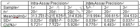
Intra-assay Precision (Precision within an assay): CV%<8%

Three samples of known concentration were tested twenty times on one plate to assess.

Inter-assay Precision (Precision between assays): CV%<10%

Three samples of known concentration were tested in twenty assays to assess.
 

Linearity


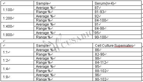
To assess the linearity of the assay, samples were spiked with high concentrations of mouse VCAM-1/CD106 in various matrices and diluted with the Sample Diluent to produce samples with values within the dynamic range of the assay.
 

Typical Data


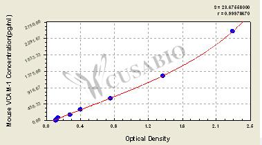
These standard curves are provided for demonstration only. A standard curve should be generated for each set of samples assayed.


Troubleshooting and FAQs
Images
  • Mouse Vascular cell adhesion molecule 1,VCAM-1 ELISA kit
  • Mouse Vascular cell adhesion molecule 1,VCAM-1 ELISA kit
Description

This mouse VCAM1 ELISA kit employs the quantitative sandwich enzyme immunoassay technique to measure the levels of mouse VCAM1 in the serum, plasma, cell culture supernates, or tissue homogenates. Antibody specific for VCAM1 has been pre-coated onto the microplate. Standards and samples are pipetted into the wells and any VCAM1 present is bound by the immobilized antibody. After removing any unbound substances, a biotin-conjugated VCAM1 antibody is added to the wells. After washing, avidin conjugated HRP is added to the wells, forming an antibody-antigen-enzyme-labeled antibody complex. Following a wash to remove any unbound HRP-avidin, the TMB substrate solution is added to the wells, and the color develops into blue. The color changes from blue to yellow after the addition of stop solution into the wells. The color intensity is in proportion to the amount of VCAM1 bound in the initial step.

VCAM1 is inducibly and mainly expressed in endothelial cells. As a cell adhesion molecule, VCAM1 contributes to modulating inflammation-related vascular adhesion and the transendothelial migration of leukocytes, such as macrophages and T cells. VCAM1 is closely linked to the occurrence and development of inflammatory diseases, including rheumatoid arthritis, asthma, and transplant rejection. Many studies have shown that VCAM1 is expressed in different cancer types such as breast, renal, and gastric cancers. High expression of VCAM1 has been found in breast cancer and gastric cancer and is associated with tumor angiogenesis and metastasis.

Customer Reviews and Q&A

 Customer Reviews

Target Background

Function(From Uniprot)
Important in cell-cell recognition. Appears to function in leukocyte-endothelial cell adhesion. Interacts with integrin alpha-4/beta-1 (ITGA4/ITGB1) on leukocytes, and mediates both adhesion and signal transduction. The VCAM1/ITGA4/ITGB1 interaction may play a pathophysiologic role both in immune responses and in leukocyte emigration to sites of inflammation.
Subcellular Location
[Isoform 1]: Cell membrane; Single-pass type I membrane protein.; [Isoform 2]: Cell membrane; Lipid-anchor, GPI-anchor.
Tissue Specificity
Expressed on inflamed vascular endothelium, as well as on macrophage-like and dendritic cell types in both normal and inflamed tissue. Expressed in the bone marrow.
Database Links
icon of phone
Call us
301-363-4651 (Available 9 a.m. to 5 p.m. CST from Monday to Friday)
icon of address
Address
No. 269, Shendun 5th Road, Donghu Hi-Tech Development Area, Hubei Province, 430206, P.R.China
icon of social media
Join us with

Subscribe newsletter

Leave a message

* To protect against spam, please pass the CAPTCHA test below.
CAPTCHA verification
© 2007-2025 CUSABIO TECHNOLOGY LLC All rights reserved. 鄂ICP备15011166号-1
Select 0 Products