* Sample kit cost can be deducted as a $30 credit for each 96-assay kit of the same analyte and brand you subsequently purchase within six months until depleted. More details >>
Interested in a trial size? Please leave a message below.


| Intra-assay Precision (Precision within an assay): CV%<8% | ||||||
| Three samples of known concentration were tested twenty times on one plate to assess. | ||||||
| Inter-assay Precision (Precision between assays): CV%<10% | ||||||
| Three samples of known concentration were tested in twenty assays to assess. | ||||||
| To assess the linearity of the assay, samples were spiked with high concentrations of mouse IL-18 in various matrices and diluted with the Sample Diluent to produce samples with values within the dynamic range of the assay. | ||||||
| Sample | Serum(n=4) | |||||
| 1:1 | Average % | 88 | ||||
| Range % | 82-94 | |||||
| 1:2 | Average % | 97 | ||||
| Range % | 91-103 | |||||
| 1:4 | Average % | 93 | ||||
| Range % | 88-99 | |||||
| 1:8 | Average % | 94 | ||||
| Range % | 90-100 | |||||
| The recovery of mouse IL-18 spiked to levels throughout the range of the assay in various matrices was evaluated. Samples were diluted prior to assay as directed in the Sample Preparation section. | ||||||
| Sample Type | Average % Recovery | Range | ||||
| Serum (n=5) | 95 | 90-100 | ||||
| EDTA plasma (n=4) | 99 | 94-107 | ||||
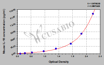
| These standard curves are provided for demonstration only. A standard curve should be generated for each set of samples assayed. | |||||||||||||||||||||||||||||||||||||||||||||||||||||||||||||||
|
|||||||||||||||||||||||||||||||||||||||||||||||||||||||||||||||
This mouse IL-18 ELISA kit uses the quantitative sandwich enzyme immunoassay technique to measure the levels of mouse IL-18 in the samples, including serum, plasma, cell culture supernates, and tissue homogenates. Antibody specific for IL-18 has been pre-coated onto the microplate. Standards and samples are pipetted into the wells and any IL-18 present is bound by the immobilized antibody. After removing any unbound substances, a biotin-conjugated IL-18 antibody is added to the wells. After washing, avidin conjugated Horseradish Peroxidase (HRP) is added to the wells, forming an antibody-antigen-enzyme-labeled antibody complex. Following a wash to remove any unbound HRP-avidin, the TMB substrate solution is added to the wells and the color develops into blue. The color changes from blue to yellow after adding the stop solution to the wells. The color intensity is proportional to the amount of IL-18 bound in the initial step.
IL-18 is a powerful pro-inflammatory cytokine involved in the regulation of both innate and acquired immune responses. It plays a key role in autoimmune, inflammatory, and infectious diseases. IL‐18 binds to IL‐18Rα and IL‐18Rβ, activating NF‐κB and inducing inflammatory mediators such as adhesion molecules, chemokines, and cellular adhesion molecules. In certain cell types, IL18 also induces MAPK and PI3K/ATK signaling pathways resulting in the generation and release of pro-inflammatory cytokines. IL-18 is associated with obesity, insulin resistance, hypertension, and dyslipidemia. High expression of IL-18 has been found in many cancer types, including colorectal cancer, multiple myeloma, and pancreatic cancer, and has been associated with cell migration, invasion, proliferation, and metastasis of tumor cells.
Sample species :
Sample type:
Review:
By Anonymous
Sample species :
Sample type:
Review:
By Anonymous
Sample species :
Sample type:
Review:
By Anonymous
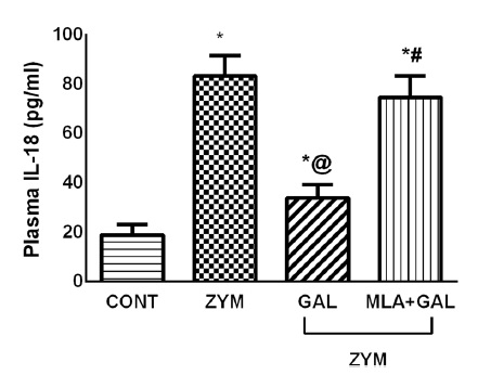
Sample species :
Sample type:
Review:
By Anonymous
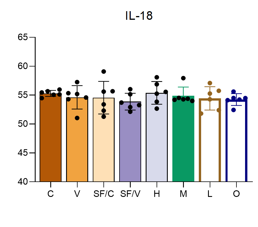
Sample species :
Sample type:
Sample dilution:
Review:
By Anonymous





