
Call us
301-363-4651 (Available 9 a.m. to 5 p.m. CST from Monday to Friday)

| Application | Recommended Dilution |
|---|---|
| WB | 1:500-1:5000 |
| IF | 1:100-1:300 |
| IP | 1:1000-1:1500 |
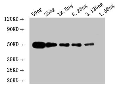
WB: Mouse anti Myc-tagged fusion protein Monoclonal antibody at 1.6μg/ml
Lane 1: Recombinant Myc-tagged fusion protein at 50ng
Lane 2: Recombinant Myc-tagged fusion protein at 25ng
Lane 3: Recombinant Myc-tagged fusion protein at 12.25ng
Lane 4: Recombinant Myc-tagged fusion protein at 6.25ng
Lane 5: Recombinant Myc-tagged fusion protein at 3.125ng
Lane 6: Recombinant Myc-tagged fusion protein at 1.5625ng
Secondary
Goat polyclonal to Mouse IgG at 1/50000 dilution
Predicted band size: 50 kd
Observed band size: 50 kd
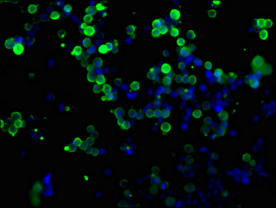
Immunofluorescence staining of transfected HEK293 cells with CSB-MA000041M0m at 1:100, counter-stained with DAPI. The cells were fixed in 4% formaldehyde, permeabilized using 0.2% Triton X-100 and blocked in 10% normal Goat Serum. The cells were then incubated with the antibody overnight at 4°C. The secondary antibody was Alexa Fluor 488-congugated AffiniPure Goat Anti-Mouse IgG(H+L).
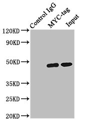
Immunoprecipitating MYC-tag with transfected HEK293
Lane 1: Mouse control IgG (1µg) instead of CSB-MA000041M0m in transfected HEK293 whole cell lysate. For western blotting, a HRP-conjugated Protein G antibody was used as the secondary antibody (1/2000)
Lane 2: CSB-MA000041M0m (6µg) + transfected HEK293 whole cell lysate (1mg)
Lane 3: Transfected HEK293 whole cell lysate (10µg)
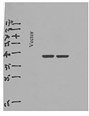
Applications :
Sample type:
Sample dilution:
Review:
By Anonymous
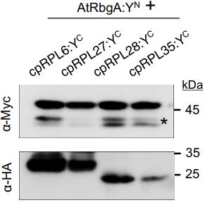
Applications :
Sample type:
Sample dilution:
Review:
By Anonymous
Applications :
Sample dilution:
Review:
By Anonymous
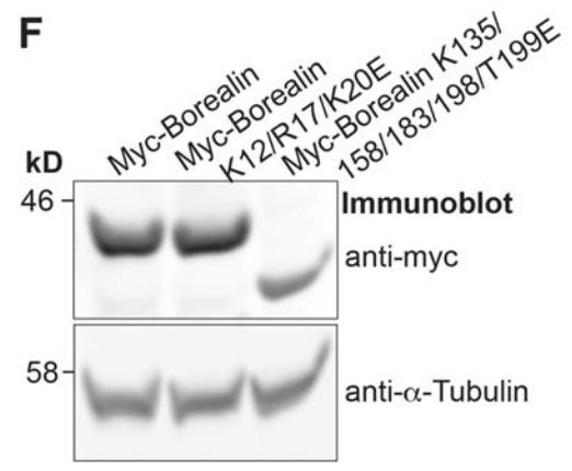
Applications :
Sample type:
Sample dilution:
Review:
By Anonymous


Email: support@cusabio.com
Distributors Worldwide



