Call us
301-363-4651 (Available 9 a.m. to 5 p.m. CST from Monday to Friday)
| Application | Recommended Dilution |
|---|---|
| WB | 1:1000-1:5000 |
| IHC | 1:20-1:500 |
| IF | 1:50-1:200 |
| IP | 1:200-1:2000 |
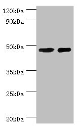
Western blot
All lanes: SMAD3 antibody at 8μg/ml
Lane 1: Jurkat whole cell lysate
Lane 2: A431 whole cell lysate
Secondary
Goat polyclonal to rabbit IgG at 1/10000 dilution
Predicted band size: 49, 44, 36, 26 kDa
Observed band size: 49 kDa
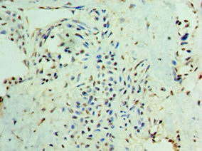
Immunohistochemistry of paraffin-embedded human breast cancer using CSB-PA021788ESR2HU at dilution of 1:100
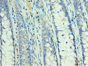
Immunohistochemistry of paraffin-embedded human colon cancer using CSB-PA021788ESR2HU at dilution of 1:100
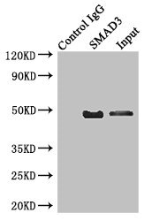
Immunoprecipitating SMAD3 in Jurkat whole cell lysate
Lane 1: Rabbit control IgG instead of (1μg) instead of CSB-PA021788ESR2HU in Jurkat whole cell lysate.
For western blotting, a HRP-conjugated anti-rabbit IgG, specific to the non-reduced form of IgG was used as the Secondary antibody (1/50000)
Lane 2: CSB-PA021788ESR2HU (4μg) + Jurkat whole cell lysate (500μg)
Lane 3: Jurkat whole cell lysate (20μg)
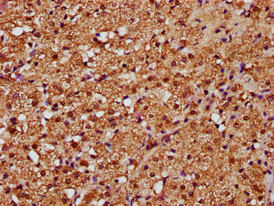
IHC image of CSB-PA021788ESR2HU diluted at 1:388 and staining in paraffin-embedded human adrenal gland tissue performed on a Leica BondTM system. After dewaxing and hydration, antigen retrieval was mediated by high pressure in a citrate buffer (pH 6.0). Section was blocked with 10% normal goat serum 30min at RT. Then primary antibody (1% BSA) was incubated at 4°C overnight. The primary is detected by a biotinylated secondary antibody and visualized using an HRP conjugated SP system.
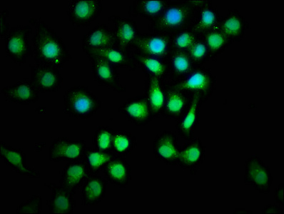
Immunofluorescence staining of A549 cells with CSB-PA021788ESR2HU at 1:129, counter-stained with DAPI. The cells were fixed in 4% formaldehyde, permeabilized using 0.2% Triton X-100 and blocked in 10% normal Goat Serum. The cells were then incubated with the antibody overnight at 4°C. The secondary antibody was Alexa Fluor 488-congugated AffiniPure Goat Anti-Rabbit IgG(H+L).


Email: support@cusabio.com
Distributors Worldwide