Call us
301-363-4651 (Available 9 a.m. to 5 p.m. CST from Monday to Friday)
| Application | Recommended Dilution |
|---|---|
| WB | 1:500-1:5000 |
| IF | 1:50-1:200 |
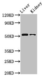
Western Blot
Positive WB detected in: Rat liver tissue, Rat kidney tissue
All lanes: PTGER4 antibody at 3.2μg/ml
Secondary
Goat polyclonal to rabbit IgG at 1/50000 dilution
Predicted band size: 54 kDa
Observed band size: 54 kDa
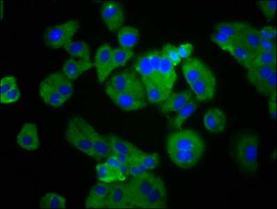
Immunofluorescence staining of HepG2 cells with CSB-PA018973LA01HU at 1:133, counter-stained with DAPI. The cells were fixed in 4% formaldehyde, permeabilized using 0.2% Triton X-100 and blocked in 10% normal Goat Serum. The cells were then incubated with the antibody overnight at 4°C. The secondary antibody was Alexa Fluor 488-congugated AffiniPure Goat Anti-Rabbit IgG(H+L).


Email: support@cusabio.com
Distributors Worldwide