Call us
301-363-4651 (Available 9 a.m. to 5 p.m. CST from Monday to Friday)
| Application | Recommended Dilution |
|---|---|
| WB | 1:200-1:2000 |
| IHC | 1:20-1:200 |
| IF | 1:50-1:200 |
| IP | 1:200-1:2000 |
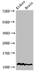
Western Blot
Positive WB detected in: Mouse kidney tissue, Mouse brain tissue
All lanes: HIST1H4A antibody at 2µg/ml
Secondary
Goat polyclonal to rabbit IgG at 1/50000 dilution
Predicted band size: 12 kDa
Observed band size: 12 kDa
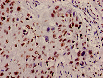
Immunohistochemistry of paraffin-embedded human cervical cancer using CSB-PA010429PA12nacHU at dilution of 1:100
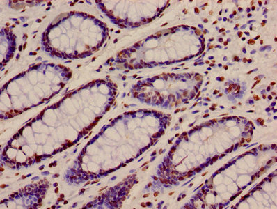
Immunohistochemistry of paraffin-embedded human colon cancer using CSB-PA010429PA12nacHU at dilution of 1:100
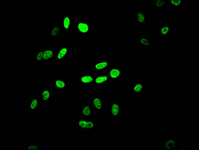
Immunofluorescent analysis of Hela cells using CSB-PA010429PA12nacHU at dilution of 1:100 and Alexa Fluor 488-congugated AffiniPure Goat Anti-Rabbit IgG(H+L)
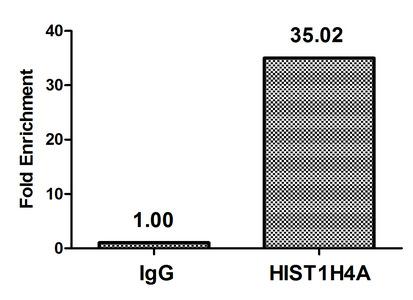
Chromatin Immunoprecipitation Hela (4*106) were treated with Micrococcal Nuclease, sonicated, and immunoprecipitated with 8µg anti-HIST1H4A (CSB-PA010429PA12nacHU) or a control normal rabbit IgG. The resulting ChIP DNA was quantified using real-time PCR with primers against the β-Globin promoter.
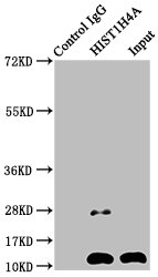
Immunoprecipitating HIST1H4A in Mouse brain tissue
Lane 1: Rabbit control IgG instead of CSB-PA010429PA12nacHU in Mouse brain tissue. For western blotting, a HRP-conjugated Protein G antibody was used as the secondary antibody (1/2000)
Lane 2: CSB-PA010429PA12nacHU (5µg) + Mouse brain tissue (500µg)
Lane 3: Mouse brain tissue (20µg)


Email: support@cusabio.com
Distributors Worldwide