
Call us
301-363-4651 (Available 9 a.m. to 5 p.m. CST from Monday to Friday)

| Application | Recommended Dilution |
|---|---|
| WB | 1:100-1:1000 |
| ICC | 1:10-1:100 |
| IF | 1:1-1:10 |
| IP | 1:200-1:2000 |
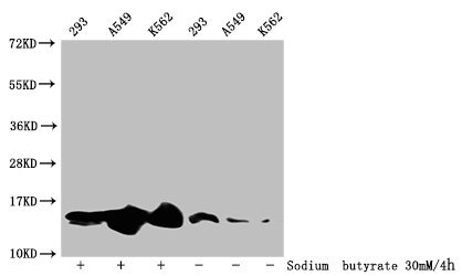
Western Blot
Detected samples: 293 whole cell lysate, A549 whole cell lysate, K562 whole cell lysate; Untreated (-) or treated (+) with 30mM sodium butyrate for 4h
All lanes: HIST1H2BC antibody at 1:100
Secondary
Goat polyclonal to rabbit IgG at 1/50000 dilution
Predicted band size: 14 kDa
Observed band size: 14 kDa
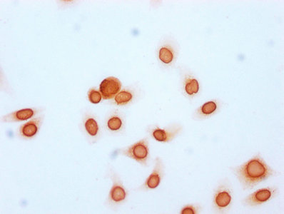
Immunocytochemistry analysis of CSB-PA010403OA20acHU diluted at 1:15 and staining in Hela cells (treated with 30mM sodium butyrate for 4h) performed on a Leica BondTM system. The cells were fixed in 4% formaldehyde, permeabilized using 0.2% Triton X-100 and blocked with 10% normal goat serum 30min at RT. Then primary antibody (1% BSA) was incubated at 4°C overnight. The primary is detected by a biotinylated secondary antibody and visualized using an HRP conjugated SP system.
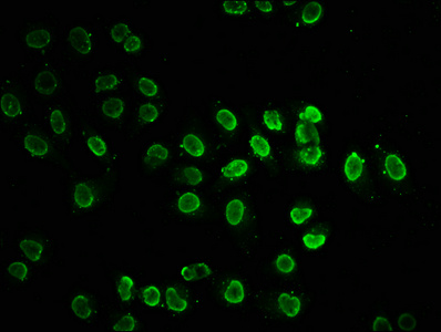
Immunofluorescence staining of Hela cells (treated with 30mM sodium butyrate for 4h) with CSB-PA010403OA20acHU at 1:7.5, counter-stained with DAPI. The cells were fixed in 4% formaldehyde, permeabilized using 0.2% Triton X-100 and blocked in 10% normal Goat Serum. The cells were then incubated with the antibody overnight at 4°C. The secondary antibody was Alexa Fluor 488-congugated AffiniPure Goat Anti-Rabbit IgG(H+L).
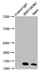
Immunoprecipitating HIST1H2BC in A549 whole cell lysate (treated with 30mM sodium butyrate for 4h)
Lane 1: Rabbit control IgG instead of CSB-PA010403OA20acHU in A549 whole cell lysate (treated with 30mM sodium butyrate for 4h). For western blotting, a HRP-conjugated Protein G antibody was used as the secondary antibody (1/2000)
Lane 2: CSB-PA010403OA20acHU (5µg) + A549 whole cell lysate (treated with 30mM sodium butyrate for 4h) (500µg)
Lane 3: A549 whole cell lysate (treated with 30mM sodium butyrate for 4h) (20µg)
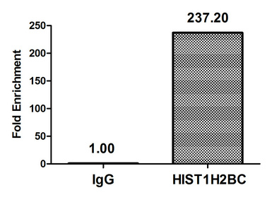
Chromatin Immunoprecipitation Hela (106, treated with 30mM sodium butyrate for 4h) were treated with Micrococcal Nuclease, sonicated, and immunoprecipitated with 5µg anti-HIST1H2BC (CSB-PA010403OA20acHU) or a control normal rabbit IgG. The resulting ChIP DNA was quantified using real-time PCR with primers against the β-Globin promoter.


Email: support@cusabio.com
Distributors Worldwide



