
Call us
301-363-4651 (Available 9 a.m. to 5 p.m. CST from Monday to Friday)

| Application | Recommended Dilution |
|---|---|
| WB | 1:2000-1:80000 |
| IHC | 1:50-1:200 |
| IF | 1:50-1:200 |
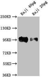
Western Blot
Positive WB detected in: Raji whole cell lysate
All lanes: CD19 antibody at 1:2000
Secondary
Goat polyclonal to Mouse IgG at 1/10000 dilution
Predicted band size: 61 kDa
Observed band size: 95 kDa
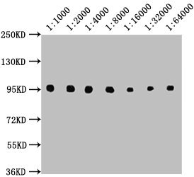
Western Blot
Positive WB detected in: Raji whole cell lysate
All lanes: CD19 antibody at 1:1000, 1:2000, 1:4000, 1:8000, 1:16000, 1:32000, 1:64000
Secondary
Goat polyclonal to Mouse IgG at 1/10000 dilution
Predicted band size: 61 kDa
Observed band size: 95 kDa
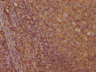
IHC image of CSB-MA004888A0m diluted at 1:100 and staining in paraffin-embedded human tonsil tissue performed on a Leica BondTM system. After dewaxing and hydration, antigen retrieval was mediated by high pressure in a citrate buffer (pH 6.0). Section was blocked with 10% normal goat serum 30min at RT. Then primary antibody (1% BSA) was incubated at 4°C overnight. The primary is detected by a biotinylated secondary antibody and visualized using an HRP conjugated SP system.
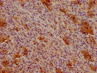
IHC image of CSB-MA004888A0m diluted at 1:100 and staining in paraffin-embedded human spleen tissue performed on a Leica BondTM system. After dewaxing and hydration, antigen retrieval was mediated by high pressure in a citrate buffer (pH 6.0). Section was blocked with 10% normal goat serum 30min at RT. Then primary antibody (1% BSA) was incubated at 4°C overnight. The primary is detected by a biotinylated secondary antibody and visualized using an HRP conjugated SP system.
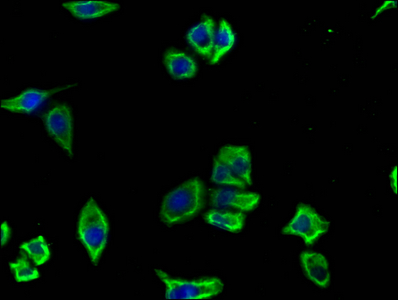
Immunofluorescence staining of Hela cells with CSB-MA004888A0m at 1:100, counter-stained with DAPI. The cells were blocked in 10% normal Goat Serum and then incubated with the primary antibody overnight at 4°C. The secondary antibody was Alexa Fluor 488-congugated AffiniPure Goat Anti-Mouse IgG(H+L).
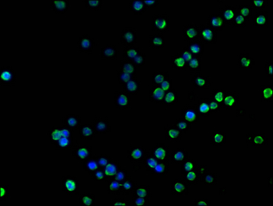
Immunofluorescence staining of Raji cells with CSB-MA004888A0m at 1:100, counter-stained with DAPI. The cells were blocked in 10% normal Goat Serum and then incubated with the primary antibody overnight at 4°C. The secondary antibody was Alexa Fluor 488-congugated AffiniPure Goat Anti-Mouse IgG(H+L).
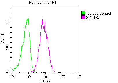
Overlay histogram showing Raji cells stained with CSB-MA004888A0m (red line) at 1:100. The cells were incubated in 1x PBS /10% normal goat serum to block non-specific protein-protein interactions followed by primary antibody for 1 h at 4°C. The secondary antibody used was FITC goat anti-mouse IgG(H+L) at 1/200 dilution for 1 h at 4°C. Isotype control antibody (green line) was used under the same conditions. Acquisition of >10,000 events was performed.
The CD19 Monoclonal Antibody is a murine non-conjugated monoclonal IgG1 antibody, which is reactive to the human CD19 on its 20-291aa region (extracellular region). This antibody was purified using protein A, with a purity greater than 95%. It can be used in ELISA, Western Blot (WB), Immunohistochemistry (IHC), Immunofluorescence (IF), and Flow Cytometry (FC) for studies related to immunology, like antigen response, B-cell maturity cycle and immunoglobulin production. The human CD19 protein is a B-lymphocyte antigen that is a coreceptor for the B-cell antigen receptor complex, decreasing the threshold for B-cell activation and B-cell response against antigens. It’s vital for normal B-cell differentiation and proliferation in response to antigen challenge and to maintain healthy levels of serum immunoglobulins. It’s also related to a medical condition called Immunodeficiency, common variable, 3 (CVID3). In this disease, the CD19 is truncated or mutated, causing B-cell faulty differentiation and impaired immunoglobulin secretion, which leads to symptoms like antibody deficiency (hypogammaglobulinemia) and recurrent bacterial infections.


Email: support@cusabio.com
Distributors Worldwide



