
Call us
301-363-4651 (Available 9 a.m. to 5 p.m. CST from Monday to Friday)

| Application | Recommended Dilution |
|---|---|
| WB | 1:1000-1:5000 |
| IHC | 1:50-1:200 |
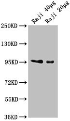
Western Blot
Positive WB detected in: Raji whole cell lysate
All lanes: CD19 antibody at 1:2000
Secondary
Goat polyclonal to Mouse IgG at 1/10000 dilution
Predicted band size: 61 kDa
Observed band size: 95 kDa
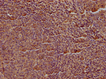
IHC image of CSB-MA004888A1m diluted at 1:100 and staining in paraffin-embedded human tonsil tissue performed on a Leica BondTM system. After dewaxing and hydration, antigen retrieval was mediated by high pressure in a citrate buffer (pH 6.0). Section was blocked with 10% normal goat serum 30min at RT. Then primary antibody (1% BSA) was incubated at 4°C overnight. The primary is detected by a biotinylated secondary antibody and visualized using an HRP conjugated SP system.
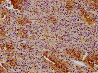
IHC image of CSB-MA004888A1m diluted at 1:100 and staining in paraffin-embedded human spleen tissue performed on a Leica BondTM system. After dewaxing and hydration, antigen retrieval was mediated by high pressure in a citrate buffer (pH 6.0). Section was blocked with 10% normal goat serum 30min at RT. Then primary antibody (1% BSA) was incubated at 4°C overnight. The primary is detected by a biotinylated secondary antibody and visualized using an HRP conjugated SP system.
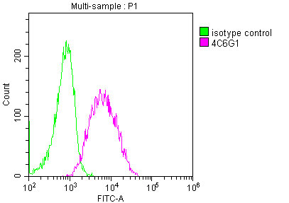
Overlay histogram showing Raji cells stained with CSB-MA004888A1m (red line) at 1:50. The cells were incubated in 1x PBS /10% normal goat serum to block non-specific protein-protein interactions followed by primary antibody for 1 h at 4°C. The secondary antibody used was FITC goat anti-mouse IgG(H+L) at 1/200 dilution for 1 h at 4°C. Isotype control antibody (green line) was used under the same conditions. Acquisition of >10,000 events was performed.
The production of the CD19 monoclonal antibody began with immunization of mouse & isolation of splenocytes. A Recombinant Human CD19 protein (20-291AA) was injected into the mouse and later its blood was screened for the next step. The splenocytes were then isolated for in vitro hybridoma production. At the same time, the myeloma cells were prepared. Myeloma cells and isolated splenocytes were fused together to form hybridomas with hybridoma technology. Next, hybridomas were screened and cloned. Finally, the CD19 monoclonal antibody was produced and validated in ELISA, WB, IHC, FC.


Email: support@cusabio.com
Distributors Worldwide



