* Sample kit cost can be deducted as a $30 credit for each 96-assay kit of the same analyte and brand you subsequently purchase within six months until depleted. More details >>
Interested in a trial size? Please leave a message below.


| Intra-assay Precision (Precision within an assay): CV%<8% | ||||||
| Three samples of known concentration were tested twenty times on one plate to assess. | ||||||
| Inter-assay Precision (Precision between assays): CV%<10% | ||||||
| Three samples of known concentration were tested in twenty assays to assess. | ||||||
| To assess the linearity of the assay, samples were spiked with high concentrations of mouse α-MSH in various matrices and diluted with the Sample Diluent to produce samples with values within the dynamic range of the assay. | ||||||
| Sample | Serum(n=4) | |||||
| 1:1 | Average % | 89 | ||||
| Range % | 86-95 | |||||
| 1:2 | Average % | 92 | ||||
| Range % | 88-99 | |||||
| 1:4 | Average % | 94 | ||||
| Range % | 90-99 | |||||
| 1:8 | Average % | 93 | ||||
| Range % | 87-98 | |||||
| The recovery of mouse α-MSH spiked to levels throughout the range of the assay in various matrices was evaluated. Samples were diluted prior to assay as directed in the Sample Preparation section. | ||||||
| Sample Type | Average % Recovery | Range | ||||
| Serum (n=5) | 93 | 89-97 | ||||
| EDTA plasma (n=4) | 95 | 91-99 | ||||
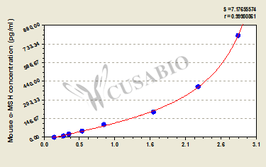
| These standard curves are provided for demonstration only. A standard curve should be generated for each set of samples assayed. | |||||||||||||||||||||||||||||||||||||||||||||||||||||||||||||||
| |||||||||||||||||||||||||||||||||||||||||||||||||||||||||||||||
α-Melanocyte Stimulating Hormone (α-MSH) is a peptide hormone derived from pro-opiomelanocortin (POMC) that plays key roles in melanogenesis, appetite regulation, and energy homeostasis. This neuropeptide acts through melanocortin receptors to stimulate melanin production in melanocytes and influences feeding behavior through hypothalamic pathways. α-MSH also has anti-inflammatory properties and participates in various physiological processes including circadian rhythm regulation and sexual behavior, making it an important target for research in endocrinology, neuroscience, and metabolic studies.
The Mouse α-Melanocyte Stimulating Hormone (α-MSH) ELISA Kit (CSB-E15876m) uses a quantitative sandwich measurement principle for detecting α-MSH in mouse samples. This assay works with serum, plasma, and cell culture supernatants with a detection range of 12.5-800 pg/mL and sensitivity of 3.12 pg/mL. The protocol requires 50-100 μL sample volume and can be completed within 1-5 hours. Detection occurs at 450 nm wavelength.
Application Examples
Note: The following application examples are drawn from a selection of publications citing this product. For additional applications, please refer to the full list of references in the "Citations" section.
This ELISA kit has been used in research examining melanocortin signaling pathways and their roles in metabolic regulation and pigmentation processes. Studies have applied the kit to measure α-MSH levels in various biological samples including serum and tissue homogenates from mouse models.
• Metabolic research: Measuring α-MSH concentrations in serum samples from mouse studies examining adrenocorticotropic hormone and melanocyte-stimulating hormone pathways
• Pigmentation studies: Determining α-MSH content in skin tissue homogenates alongside other melanogenesis-related proteins to study pigment regulation mechanisms
• Comparative studies: Measuring α-MSH levels in healthy versus disease state samples to explore potential biomarker associations
• Multi-analyte profiling: Combining with other melanocortin pathway components including POMC, MC1R, MITF, and pigmentation enzymes in comprehensive tissue analysis





